Journal of Occupational
Health and Epidemiology

Rafsanjan university Of medical sciences
Volume 10, Issue 4 (Autumn 2021)
J Occup Health Epidemiol 2021, 10(4): 209-216 |
Back to browse issues page


Download citation:
BibTeX | RIS | EndNote | Medlars | ProCite | Reference Manager | RefWorks
Send citation to:


BibTeX | RIS | EndNote | Medlars | ProCite | Reference Manager | RefWorks
Send citation to:
Alian Samakkhah S, Tooryan F, Hushmandi K, Partovi R. Prevalence of Parasitic Infections in the Liver of Slaughtered Animals and Its Economic Losses in Modern Slaughterhouses of Mazandaran Province during 2018-2019; A Retrospective Study. J Occup Health Epidemiol 2021; 10 (4) :209-216
URL: http://johe.rums.ac.ir/article-1-465-en.html
URL: http://johe.rums.ac.ir/article-1-465-en.html
Related article in
Google Scholar
Google Scholar
Similar articles


1- Assistant Prof., Dept. of Food Hygiene, Faculty of Veterinary Medicine, Amol University of Special Modern Technologies, Amol, Iran. , Shohre.alian@ut.ac.ir
2- Assistant Prof., Dept. of Food Hygiene, Faculty of Veterinary Medicine, Amol University of Special Modern Technologies, Amol, Iran.
3- PhD Candidate in Epidemiology, Dept. of Food Hygiene and Quality Control, Division of Epidemiology & Zoonoses, Faculty of Veterinary Medicine, University of Tehran, Tehran, Iran
Article history

Received: 2021/08/29
Accepted: 2021/10/13
ePublished: 2021/12/25
Accepted: 2021/10/13
ePublished: 2021/12/25
Subject:
Epidemiology
Full-Text [PDF 400 kb]
(700 Downloads)
| Abstract (HTML) (1378 Views)
Full-Text: (11 Views)
Prevalence of Parasitic Infections in the Liver of Slaughtered Animals and Its Economic Losses in Modern Slaughterhouses of Mazandaran Province during 2018-2019; A Retrospective Study
Shohreh Alian Samakkhah1*, Fahimeh Tooryan1, Kiavash Hushmandi2, Razieh Partovi1
1. Assistant Prof., Dept. of Food Hygiene, Faculty of Veterinary Medicine, Amol University of Special Modern Technologies, Amol, Iran.
2. PhD Candidate in Epidemiology, Dept. of Food Hygiene and Quality Control, Division of Epidemiology & Zoonoses, Faculty of Veterinary Medicine, University of Tehran, Tehran, Iran.
* Corresponding author: Shohreh Alian Samakkhah; E-mail: Shohre.alian@ut.ac.ir
Abstract
Background: Regarding the consequences of parasitic infections, this study aimed to determine the prevalence and types of parasitic infections in the liver of slaughtered animals and their economic losses.
Methods and Methods: This descriptive cross-sectional study was performed on 510,802 and 501,108 head of livestock, respectively, in 2018 and 2019, including different species of cattle, sheep, and goats in the modern slaughterhouse of Mazandaran province. All recorded livers were evaluated using macroscopic, observational, and palpation methods. Data analysis was performed using SPSS software and the Chi-square test.
Results: The prevalence of parasitic infection of hydatid cyst in 2018 was 6.37% in sheep (liver), 2.40% in cattle, and 1.90% in goats; further, in 2019, it was 8.06% in sheep, 2.83% in goats, and 1.35% in cattle. According to the results, the prevalence of hydatid cyst was significantly higher in sheep liver than those in cattle and goats. After hydatidosis, fasciolosis was most prevalent in the liver of slaughtered animals. The highest seasonal prevalence of parasitic infections in the liver was observed in spring and summer, respectively. The average overall direct economic loss during the years of the study was estimated to be 82,362,000,000 Rial, equivalent to 588,300 USD.
Conclusions: The rate of contamination of slaughtered animals with hydatid cysts and Fasciola is high due to the zoonotic nature of these parasites, and the damage caused by the extermination of infected areas imposes high costs on society; thus, preventive measures should be taken in livestock in this area.
Keywords: Liver, Parasitic Infection, Slaughtered Animals, Livestock
Introduction
Evaluating the health and economic importance of each disease is the first step in determining priorities and control or prevention programs. Many animal diseases are common between humans and animals [1]. Due to the significant economic losses of liver parasites in the public health and livestock industry, caused diseases are considered as one of the important health and economic problems. Hydatid cyst is one of the most common human and animal diseases globally, which is caused by the larval stages of the Echinococcus granulosus parasite [2]. In the evolutionary cycle of this parasite, domestic and wild carnivores, especially dogs, are the final hosts, while herbivores and humans are the intermediate hosts. Deployment of its larvae in organs such as the liver and lungs and sometimes the brain, heart, and spinal cord of intermediate hosts, including humans, cause hydatidosis [3]. Humans are accidentally infected with hydatid cysts through the consumption of contaminated vegetables, contact with dogs, and geophagia [4]. The importance of the disease in humans is due to involving the sensitive and vital organs of the body, such as the liver and lungs, and in herbivores due to the substantial damage to the economy and livestock. The definitive diagnosis of hydatid cyst in cattle is made by necropsy or post-mortem inspection [1]. Fasciola is another liver parasite that lives in the bile ducts and gallbladder. Fasciolosis causes disease in humans and animals. Lymnaea snails are the intermediate host of this parasite, while ruminants are its main host [5]. Ruminants become infected by eating aquatic plants infected with the parasite. In the meantime, humans can accidentally get infected by eating these plants [6]. Dicrocoeliosis is another ruminant parasitic infection caused by different species of Dicrocoelium. The bile ducts of domestic and wild ruminants are infected with Dicrocoelium. Ruminants are the definitive hosts of this parasite, which become infected by eating the ant intermediate host containing the metacercial form of Dicrocoelium. Humans, dogs, and horses are accidentally infected with this parasite. The symptoms of this parasite are far less than those of fasciolosis. The economic and veterinary importance of Dicrocoeliosis is due to direct damage to the liver, leading to the removal of the liver in the slaughterhouse [7]. Cysticercosis is caused every year by the Taenia saginata parasite, which enters the human body through eating undercooked beef. Cysts of this parasite are mostly found in different anatomical areas, such as the liver, mesentery, omentum, and peritoneum. In sheep liver, there are usually several migration routes in reddish-gray or white-gray colors [8]. In Iran, hydatidosis is common in domestic animals, with a prevalence between 1.5 to 64% in sheep,
cattle, goats, buffalo, and camels [9]. The prevalence of fasciolosis and Dicrocoeliosis is between 2.4% and 82% for Fasciola hepatica and 2.5% to 15.6% for Dicrocoelium dendriticum [10].
Due to the significant economic losses of liver parasites in the public health and livestock industry, such diseases are considered as one of the important health and economic problems [11]. Although there may be errors in recording information in slaughterhouses, it can be stated that slaughterhouses are the only and best place to estimate the extent of contamination because diseases such as hydatidosis, fasciolosis, dicrocoeliasis, and liver lesions are mainly visible and recognizable to the naked eye; accordingly, infected organs are seized and removed from the consumption cycle [12]. Numerous studies have been conducted in different provinces and animal species in the country [13, 14].
Due to the lack of studies on the status of parasitic infections of slaughtered livestock in Mazandaran province and the high livestock population and humidity due to the Caspian Sea, livestock in this province is prone to parasitic infections. Therefore, given the consequences of parasitic infections, this study aims to determine the prevalence and types of parasitic infections in the liver of slaughtered animals in modern slaughterhouses in Mazandaran province and their economic losses during 2018-2019.
Materials and Methods
Mazandaran province, with an area of 23842 square kilometers, is located in northern Iran, which has a humid and temperate climate in terms of natural conditions due to the sea, Alborz Mountains, and forests. It is one of the most important provinces in Iran in the field of cattle, sheep, and goats. The average temperature of this province is 25 degrees Celsius in summer and 9 degrees Celsius in winter [15]. Figure 1 shows the location of the study area.
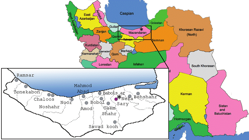
Fig. 1. Location of the study area
This study was descriptive and cross-sectional, being conducted for two years from the first of April 2018 to the end of March 2019. Sampling was done by the census method; thus, the sample size included all slaughtered animals during the study period. For this study, data related to the total slaughtered livestock (cattle, sheep, goats) in the active modern slaughterhouses in the province, under the supervision of the General Veterinary Office of Mazandaran Province, were collected. This data included daily recorded information about the number of seized livers. Diagnosis of lesions in the liver of cattle was made by observing, palpating, and incising the affected areas by a veterinarian and slaughterhouse inspectors, based on the meat inspection protocol of the Food and Agriculture Organization of the United Nations. This protocol states that visual examination with palpation should be made for abscesses, parasitic infections such as hydatid cysts, and fascioliasis. The incision should be made on the gastric surface of the liver (in bovines, an incision at the base of the caudate lobe) to examine the bile ducts. Where necessary for a diagnosis, incise as necessary into the bile ducts and liver substance [16].
Based on condemned livers due to parasitic infections, the direct economic loss was calculated as the following procedure:
Formula 1.
DFL= CL × P × W [17]
DFL: Direct Financial Loss,
CL: Number of Condemned Livers due to parasitic infections,
P: Average Liver Price (Rial/Kg),
W: Average Liver Weight (Kg).
The average weights of cattle and ovine liver (W) were determined by weighing 30 livers of each group of animals of different ages. The average weights were calculated as 1 kg and 5 kg for ovine
and cattle livers in this region, respectively. The average sell prices (P) for each kilogram of ovine and cattle liver were 700,000 and 450,000 Rial, respectively, acquired by interviewing local butchers in different areas in Mazandaran province for two years [18].
Descriptive results were calculated as absolute frequency, relative frequency, disease prevalence. Moreover, the differences in the prevalence of parasitic infections between different animal species, seasons, and years studied were analyzed by the Chi-square test. Economic losses due to liver condemnation during years of study were calculated. Data analysis was performed by SPSS software version 25 (SPSS Inc., Chicago, IL, USA). In all analyzes, (P <0.05) were considered as statistically [19]. The Ethics Committee of Amol University of special modern technologies, Iran, approved this study (IR.AUSMT.REC.1399.173).
Results
In 2018, a total of 510,802 livestock were slaughtered, and 5,303 cattle livers, 48,001 sheep livers, and 5708 goat livers were seized; the reasons for their seizure were parasitic infections, being 3766 (72.02%), 38819 (80.87%), and 5391 (94.44%), respectively. As shown in Table 1, the highest prevalence of parasitic infections in the liver was related to the hydatid cyst. The prevalence of hydatid cyst infection was 6.37% in sheep, 2.40% in cattle, and 1.90% in goats. The prevalence of hydatid cyst was significantly higher in sheep liver than that of cattle and goats (P = 0.001). The other parasitic infections in the seized livers of cattle, sheep, and goats included Fasciola, Dicrocoelium, and Cysticercus, the prevalence of which is presented in Table 1. According to the results, the prevalence of parasitic infections (Fasciola and Dicrocoelium) in sheep was higher than in cattle and goats (P = 0.001). Infection with Cysticercus was the same in the liver of the studied animals (P = 0.08).
Table 1. The prevalence of different parasitic infections in the liver of slaughtered animals based on type in modern slaughterhouses of Mazandaran province during 2018
Shohreh Alian Samakkhah1*, Fahimeh Tooryan1, Kiavash Hushmandi2, Razieh Partovi1
1. Assistant Prof., Dept. of Food Hygiene, Faculty of Veterinary Medicine, Amol University of Special Modern Technologies, Amol, Iran.
2. PhD Candidate in Epidemiology, Dept. of Food Hygiene and Quality Control, Division of Epidemiology & Zoonoses, Faculty of Veterinary Medicine, University of Tehran, Tehran, Iran.
* Corresponding author: Shohreh Alian Samakkhah; E-mail: Shohre.alian@ut.ac.ir
Abstract
Background: Regarding the consequences of parasitic infections, this study aimed to determine the prevalence and types of parasitic infections in the liver of slaughtered animals and their economic losses.
Methods and Methods: This descriptive cross-sectional study was performed on 510,802 and 501,108 head of livestock, respectively, in 2018 and 2019, including different species of cattle, sheep, and goats in the modern slaughterhouse of Mazandaran province. All recorded livers were evaluated using macroscopic, observational, and palpation methods. Data analysis was performed using SPSS software and the Chi-square test.
Results: The prevalence of parasitic infection of hydatid cyst in 2018 was 6.37% in sheep (liver), 2.40% in cattle, and 1.90% in goats; further, in 2019, it was 8.06% in sheep, 2.83% in goats, and 1.35% in cattle. According to the results, the prevalence of hydatid cyst was significantly higher in sheep liver than those in cattle and goats. After hydatidosis, fasciolosis was most prevalent in the liver of slaughtered animals. The highest seasonal prevalence of parasitic infections in the liver was observed in spring and summer, respectively. The average overall direct economic loss during the years of the study was estimated to be 82,362,000,000 Rial, equivalent to 588,300 USD.
Conclusions: The rate of contamination of slaughtered animals with hydatid cysts and Fasciola is high due to the zoonotic nature of these parasites, and the damage caused by the extermination of infected areas imposes high costs on society; thus, preventive measures should be taken in livestock in this area.
Keywords: Liver, Parasitic Infection, Slaughtered Animals, Livestock
Introduction
Evaluating the health and economic importance of each disease is the first step in determining priorities and control or prevention programs. Many animal diseases are common between humans and animals [1]. Due to the significant economic losses of liver parasites in the public health and livestock industry, caused diseases are considered as one of the important health and economic problems. Hydatid cyst is one of the most common human and animal diseases globally, which is caused by the larval stages of the Echinococcus granulosus parasite [2]. In the evolutionary cycle of this parasite, domestic and wild carnivores, especially dogs, are the final hosts, while herbivores and humans are the intermediate hosts. Deployment of its larvae in organs such as the liver and lungs and sometimes the brain, heart, and spinal cord of intermediate hosts, including humans, cause hydatidosis [3]. Humans are accidentally infected with hydatid cysts through the consumption of contaminated vegetables, contact with dogs, and geophagia [4]. The importance of the disease in humans is due to involving the sensitive and vital organs of the body, such as the liver and lungs, and in herbivores due to the substantial damage to the economy and livestock. The definitive diagnosis of hydatid cyst in cattle is made by necropsy or post-mortem inspection [1]. Fasciola is another liver parasite that lives in the bile ducts and gallbladder. Fasciolosis causes disease in humans and animals. Lymnaea snails are the intermediate host of this parasite, while ruminants are its main host [5]. Ruminants become infected by eating aquatic plants infected with the parasite. In the meantime, humans can accidentally get infected by eating these plants [6]. Dicrocoeliosis is another ruminant parasitic infection caused by different species of Dicrocoelium. The bile ducts of domestic and wild ruminants are infected with Dicrocoelium. Ruminants are the definitive hosts of this parasite, which become infected by eating the ant intermediate host containing the metacercial form of Dicrocoelium. Humans, dogs, and horses are accidentally infected with this parasite. The symptoms of this parasite are far less than those of fasciolosis. The economic and veterinary importance of Dicrocoeliosis is due to direct damage to the liver, leading to the removal of the liver in the slaughterhouse [7]. Cysticercosis is caused every year by the Taenia saginata parasite, which enters the human body through eating undercooked beef. Cysts of this parasite are mostly found in different anatomical areas, such as the liver, mesentery, omentum, and peritoneum. In sheep liver, there are usually several migration routes in reddish-gray or white-gray colors [8]. In Iran, hydatidosis is common in domestic animals, with a prevalence between 1.5 to 64% in sheep,
cattle, goats, buffalo, and camels [9]. The prevalence of fasciolosis and Dicrocoeliosis is between 2.4% and 82% for Fasciola hepatica and 2.5% to 15.6% for Dicrocoelium dendriticum [10].
Due to the significant economic losses of liver parasites in the public health and livestock industry, such diseases are considered as one of the important health and economic problems [11]. Although there may be errors in recording information in slaughterhouses, it can be stated that slaughterhouses are the only and best place to estimate the extent of contamination because diseases such as hydatidosis, fasciolosis, dicrocoeliasis, and liver lesions are mainly visible and recognizable to the naked eye; accordingly, infected organs are seized and removed from the consumption cycle [12]. Numerous studies have been conducted in different provinces and animal species in the country [13, 14].
Due to the lack of studies on the status of parasitic infections of slaughtered livestock in Mazandaran province and the high livestock population and humidity due to the Caspian Sea, livestock in this province is prone to parasitic infections. Therefore, given the consequences of parasitic infections, this study aims to determine the prevalence and types of parasitic infections in the liver of slaughtered animals in modern slaughterhouses in Mazandaran province and their economic losses during 2018-2019.
Materials and Methods
Mazandaran province, with an area of 23842 square kilometers, is located in northern Iran, which has a humid and temperate climate in terms of natural conditions due to the sea, Alborz Mountains, and forests. It is one of the most important provinces in Iran in the field of cattle, sheep, and goats. The average temperature of this province is 25 degrees Celsius in summer and 9 degrees Celsius in winter [15]. Figure 1 shows the location of the study area.
Fig. 1. Location of the study area
This study was descriptive and cross-sectional, being conducted for two years from the first of April 2018 to the end of March 2019. Sampling was done by the census method; thus, the sample size included all slaughtered animals during the study period. For this study, data related to the total slaughtered livestock (cattle, sheep, goats) in the active modern slaughterhouses in the province, under the supervision of the General Veterinary Office of Mazandaran Province, were collected. This data included daily recorded information about the number of seized livers. Diagnosis of lesions in the liver of cattle was made by observing, palpating, and incising the affected areas by a veterinarian and slaughterhouse inspectors, based on the meat inspection protocol of the Food and Agriculture Organization of the United Nations. This protocol states that visual examination with palpation should be made for abscesses, parasitic infections such as hydatid cysts, and fascioliasis. The incision should be made on the gastric surface of the liver (in bovines, an incision at the base of the caudate lobe) to examine the bile ducts. Where necessary for a diagnosis, incise as necessary into the bile ducts and liver substance [16].
Based on condemned livers due to parasitic infections, the direct economic loss was calculated as the following procedure:
Formula 1.
DFL= CL × P × W [17]
DFL: Direct Financial Loss,
CL: Number of Condemned Livers due to parasitic infections,
P: Average Liver Price (Rial/Kg),
W: Average Liver Weight (Kg).
The average weights of cattle and ovine liver (W) were determined by weighing 30 livers of each group of animals of different ages. The average weights were calculated as 1 kg and 5 kg for ovine
and cattle livers in this region, respectively. The average sell prices (P) for each kilogram of ovine and cattle liver were 700,000 and 450,000 Rial, respectively, acquired by interviewing local butchers in different areas in Mazandaran province for two years [18].
Descriptive results were calculated as absolute frequency, relative frequency, disease prevalence. Moreover, the differences in the prevalence of parasitic infections between different animal species, seasons, and years studied were analyzed by the Chi-square test. Economic losses due to liver condemnation during years of study were calculated. Data analysis was performed by SPSS software version 25 (SPSS Inc., Chicago, IL, USA). In all analyzes, (P <0.05) were considered as statistically [19]. The Ethics Committee of Amol University of special modern technologies, Iran, approved this study (IR.AUSMT.REC.1399.173).
Results
In 2018, a total of 510,802 livestock were slaughtered, and 5,303 cattle livers, 48,001 sheep livers, and 5708 goat livers were seized; the reasons for their seizure were parasitic infections, being 3766 (72.02%), 38819 (80.87%), and 5391 (94.44%), respectively. As shown in Table 1, the highest prevalence of parasitic infections in the liver was related to the hydatid cyst. The prevalence of hydatid cyst infection was 6.37% in sheep, 2.40% in cattle, and 1.90% in goats. The prevalence of hydatid cyst was significantly higher in sheep liver than that of cattle and goats (P = 0.001). The other parasitic infections in the seized livers of cattle, sheep, and goats included Fasciola, Dicrocoelium, and Cysticercus, the prevalence of which is presented in Table 1. According to the results, the prevalence of parasitic infections (Fasciola and Dicrocoelium) in sheep was higher than in cattle and goats (P = 0.001). Infection with Cysticercus was the same in the liver of the studied animals (P = 0.08).
Table 1. The prevalence of different parasitic infections in the liver of slaughtered animals based on type in modern slaughterhouses of Mazandaran province during 2018
| Year | Type of infection | Absolute frequency (relative frequency) | P-value* | ||
| Cattle (71945) | Sheep (320326) | Goat (118531) | |||
| 2018 | Hydatid cyst | 1732(2.40%)a | 20432(6.37%)b | 2254(1.90%)a | 0.001 |
| Fasciola | 1187(1.64%)a | 7534(2.35%)b | 1034(0.87%)a | 0.001 | |
| Dicrocoelium | 786(1.10%)a | 10532(3.28%)b | 1993(1.68%)a | 0.001 | |
| Cysticercus | 61(0.08%)a | 312(0.10%)a | 110(0.09%)a | 0.08 | |
| Total | 3766(5.23%)a | 38819(12.11%)b | 5391(4.55%)a | 0.001 | |
* P <0.05 indicates the significant difference in each row. a-b Different letters in the same row indicate a significant level.
In 2019, a total of 501108 livestock were slaughtered, and 5868 cattle livers, 53513 sheep livers, 6820 goat livers were seized; the reason for their seizure was parasitic infections, being 3374 (57.50%), 43930 (82.09%), and 6570 (96.33%), respectively. As shown in Table 2, the highest prevalence of parasitic infections in the cattle liver was related to Fasciola, and for sheep and goats was associated with hydatid cysts. The prevalence of hydatid cyst infection was 8.06% in sheep, 2.83% in goats, and 1.35% in cattle. The prevalence of hydatid cyst was significantly higher in sheep liver than those of cattle and goats (P=0.001). The other parasitic infections in the seized livers of cattle, sheep, and goats included Fasciola, Dicrocoelium, and Cysticercus, the prevalence of which is presented in Table 2. According to the results, the prevalence of parasitic infections (Fasciola and Dicrocoelium) in sheep was significantly higher than in goats and cattle (P=0.001). Cysticercus infection in sheep and goat livers was almost the same, showing a significant decrease compared to the cattle liver (P = 0.001). In general, the prevalence of various parasitic infections in 2019 was more than in 2018.
Table 2. The prevalence of different parasitic infections in the liver of slaughtered animals based on type in modern slaughterhouses of Mazandaran province during 2019
| Year | Type of infection | Absolute frequency (Relative frequency) | P-value* | ||
| Cattle (70458) | Sheep (312811) | Goat (117839) | |||
| 2019 | Hydatid cyst | 952(1.35%)a | 25228(8.06%)b | 3341(2.83%)a | 0.001 |
| Fasciola | 1436(2.04%)a | 8128(2.60%)b | 1125(0.95%)a | 0.001 | |
| Dicrocoelium | 914(1.30%)a | 10548(3.37%)b | 2099(1.78%)a | 0.001 | |
| Cysticercus | 72(0.10%)a | 26(0.008%)b | 5(0.004%)b | 0.001 | |
| Total | 3374(4.79%)a | 43930(14.04%)b | 6570(5.58%)a | 0.001 | |
* P <0.05 indicates the significant difference in each row. a-b Different letters in the same row indicate a significant level.
The seasonal prevalence of parasitic infections in the livers of slaughtered animals in 2018 and 2019 are shown in Figures 2 and 3, respectively. The prevalence of parasitic infections in the liver in 2018 and 2019 in spring was significantly higher than in other seasons (P <0.05). The chance of parasitic infections was in spring 1.88 times higher than in winter (OR= 1.88, 95%CI= 1.83-1.93). The highest prevalence of parasitic infections in the liver after spring was observed in summer. The highest seasonal prevalence of hydatid cyst (6.17%) was seen in the liver of slaughtered animals in the 2019 spring; however, this difference was not significant compared to 2018 (P >0.05) (Figure 3). The highest seasonal prevalence of Fasciola, Dicrocoelium, and Cysticercus in the liver of slaughtered animals was in the 2018 spring (2.77%, 3.16%, and 0.16%); however, these differences were not significant compared to 2019 (P <0.05) (Figure 2).
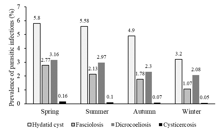
Fig. 2. Seasonal variation in the prevalence of helminths in the liver of slaughtered animals in modern slaughterhouses of Mazandaran province during 2018

Fig. 3. Seasonal variation in the prevalence of helminths in the liver of slaughtered animals in modern slaughterhouses of Mazandaran province during 2019
Fig. 2. Seasonal variation in the prevalence of helminths in the liver of slaughtered animals in modern slaughterhouses of Mazandaran province during 2018
Fig. 3. Seasonal variation in the prevalence of helminths in the liver of slaughtered animals in modern slaughterhouses of Mazandaran province during 2019
In 2018, the economic loss caused by each of the studied parasites, including hydatid cyst, fascioliasis, dicrocoeliosis, and cysticercosis, was estimated to be 19.777.200.000, 8.668.350.000, 10.36.000.000, and 432,650,000 Rial, respectively; in 2019, it was estimated to be 22.140.300.000, 9.708.100,000, 10.909.400.000, and 183.700.000 Rial, respectively
Based on the results, the average annual direct economic loss caused by parasites infections due to liver condemnation in cattle and ovine were estimated to be 39. 420.500.000 Rial in 2018 and 42.941.500.000 Rial in 2019. Furthermore, overall economic loss during the years of the study was estimated to be 82.362.000.000 Rial, equivalent to 588.300 USD.
Discussion
Parasitic infections in domestic animals are considered chronic diseases, without specific clinical symptoms, in the livestock industry, causing significant economic losses in the field. Due to the zoonotic nature of some of these diseases and the possibility of their transmission through infected meat to humans, it is essential to study the prevalence and information on appropriate strategies to reduce infection. Rejection and seizure of lung and liver are two of the most important causes of loss by hydatidosis in cattle, the economic estimate of which is very important [20].
As reported in the results of this study, the prevalence of hydatid cyst in 2018 in sheep, cattle, and goat liver was 6.37%, 2.40%, and 1.90%, respectively, while in 2019, it was 8.06%, 2.83%, and 1.35%, respectively. The prevalence of hydatid cyst was significantly higher in sheep liver than those of cattle and goats in the studied years. Also, the highest seasonal prevalence of parasitic infections in the liver was observed in spring and summer during the studied years, respectively.
In the study of Fallah et al. (2010), the infection rate was 13.7% in sheep, 16.2% in cattle, and 1.8% in goats [21]. Also, Hamzavi et al. (2016), in a study of the prevalence of hydatid cyst in slaughtered livestock in Assadabad between 2014 and 2015, reported the highest rate of infection in sheep (16.38%) and the lowest rate in goats (1.9%). Also, the highest level of infection was observed in the spring, followed by summer. The low rate of goat infection with the hydatid cyst in this study was consistent with that in the present study. This could be because the goat is less sensitive to the parasite due to the presence of protective antibodies against the parasite larvae. Also, the eating habit of goats, which prefer to feed on twigs, can be effective in reducing the incidence of hydatid cysts [22]. Ghasemian et al. (2013) examined all the livers and lungs of sheep and goats slaughtered in the Gachsaran slaughterhouse in 2012 under a macroscopic examination. A total of 11,753 livers and lungs were inspected, 1,531 of which were confiscated and taken out of the consumption cycle. According to their results, 9.99% Fasciola & 6.20% hydatid cyst in sheep liver and 27.9% Fasciola & 9.60% hydatid cyst in goat liver were observed. The prevalence of Fasciola and hydatid cyst was higher in goats than in sheep, which contradicted the present study [1]. In another aiming to determine the infection rate of slaughtered cattle in Sarpol-e-Zahab city with hydatid cyst, Cysticercus, Fasciola, and Dicrocoelium, among 1170 slaughtered cattle, 417 (35.64%) were declared infected within a year. The rates of liver infection with hydatid cyst, Fasciola, Dicrocoelium, and Cysticercus were 8.11% (95 cases), 5.47% (64 cases), 0.85% (10 cases), and 0.68% (8 cases), respectively. In the present study, the rate of infection of cattle with a high Cysticercus prevalence was reported. The lower prevalence of hydatid cysts in cattle in autumn can be related to the geographical location of the region and less rainfall in this season. In this region, unlike other areas in Iran, rainfall is usually lower in autumn; thus, the time is suitable for grazing due to the low plants, and farmers keep most of the cattle in a closed area, resulting in the low prevalence of hydatid cysts in season; autumn was less reported [23]. Majidi Rad et al. (2018) reported the Dicrocoelium infection in three provinces of Gilan, Mazandaran, and Golestan as 9.37%, 4.16%, and 1.87%, respectively [24]. Mohammadzadeh et al. (2016) reached similar results in studying the prevalence of parasitic infections in the liver and lungs of slaughtered animals in modern slaughterhouses in Fars province. They reported that the prevalence of liver hydatid cyst in slaughtered animals in Shiraz slaughterhouse was 3.44% in sheep, 3.12% in cattle, 2.94% in goats, and 2.9% in camels, respectively. Also, the prevalence of fasciolosis in sheep and cattle was 2.49% and 1.86%, respectively, while that of the Dicrocoelium parasite was 0.026% in sheep, 0.91% in cattle, and 4% in goats. According to these results, the most common infection was hydatid cyst and then fasciolosis [25]. Khanjari et al. (2015), in a study, examined the relationship of the prevalence of Cysticercus tenuicollis parasite with age, sex, season, and infected organ in slaughtered animals in Amol abattoir, Mazandaran province. They concluded that the highest prevalence of this parasite was observed in the liver of sheep and goats, with the most prevalence in spring, followed by summer [14]; this was consistent with the present study. The prevalence of liver parasites in slaughtered cattle in slaughterhouses of Lorestan province was studied by Ezzatpour et al. (2014). They reported the hydatid cyst to have the highest prevalence of parasitism in the liver of slaughtered cattle.
In the study of Kheiri et al. (2020), in the West Azerbaijan province during 2014-2019, the prevalence of hydatidosis, fasciolosis, and Dicrocoeliosis in sheep and goats was reported as 16.46%, 2.30%, and 10.50%, while in cattle, it was 15.90%, 4.70%, and 2.47%, respectively. Also, the highest prevalence of Fasciola and Dicrocoelium was observed in spring, while the highest rate of hydatidosis was in winter [26]. In our study, the highest cases of infection were related to spring, and the lowest cases were observed in winter. In Hamzavi et al. (2016), the amount of contamination in different seasons was studied, being more in spring than in other seasons [22].
In Italy (the Sardinia region), the prevalence of hydatidosis was studied by Scala et al. They reported a 75% prevalence of hydatid cyst, showing a higher prevalence of infection compared to our study [27]. Also, Odeniran et al. (2016), in a study of animals slaughtered in the Ipata slaughterhouse in Nigeria, reported the prevalence of Fasciola infection as 20.29%, 5.54%, and 2.44% in cattle, sheep, and goats, respectively. They also reported the prevalence of Dicrocoeliosis in cattle, sheep, and goats as 0.00%, 0.25%, and 0.00%, respectively. The most common organs infected by fasciolosis and Dicrocoelium were liver and bile ducts, respectively. According to these results, sheep had the most parasitic infestations and infections than cattle and goats, which was consistent with our study [28]. In examining the cattle hydatidosis prevalence while considering its economic importance in slaughtered cattle at Bahir Dar municipal abattoir, Northern Ethiopia, the total estimated annual loss due to the condemnation of offal and carcass weight loss was 1,112,769.85 Ethiopian Birr (ETB) (55,638.49 USD). Thus, efficient control measures towards hydatidosis should be applied [29]. Most African countries have records of E. granulosus. In ruminants, it has resulted in an enormous economic loss because of the condemnation of affected organs and reduction of the meat, milk, and wool production [30]. A study in Ahvaz, focusing on the economic importance of parasitic infection, reported that parasites were responsible for 54.4% of offal/carcass condemnations, with an affiliated economic loss of $1.2 million approximately. Parasites were responsible for 54.1% of the condemned organs or carcasses, with a retail value (based on market prices in 2011) of $1.148.181 (U.S.) ($137. 880 for cattle, $602,699 for sheep, $280,955 for goats, and $126,647 for buffalos) [31].
Retrospective studies of diseases encountered at abattoirs provide useful prevalence and pathology profiles, which can be used in risk assessment or future planning of control and prevention strategies. However, to achieve all these goals, rigorous and adequate meat inspection procedures, proper record of abattoir data, and, if possible, computerization of such data is necessary. It should be noted that the actual prevalence of the infection in slaughtered animals may be underestimated due to different reasons, such as potentially inadequate meat inspection, rapid slaughter rates, and unsuitable facilities. A limitation of the current study was the convenient
method used for sampling.
Conclusion
In conclusion, due to the high prevalence of hydatid cyst in Mazandaran province, this region can be considered endemic. According to the present study, the rate of contamination of slaughtered animals with hydatid cysts and Fasciola is high. One of the reasons for the high rate of parasitic infection in livestock in Mazandaran can be stray dogs and herds without health control. This indicates the potential risk of hydatid cyst disease for the health of human communities. Therefore, it is necessary for health experts and officials to take action with scientific solutions to control or prevent disease in human societies and reduce economic losses in livestock due to the reduction in livestock production and its efficiency. One of these strategies is to take anti-parasitic measures, such as using anti-parasitic drugs twice a year (early autumn and early spring) in the field.
Acknowledgement
The authors thank the colleagues of the Mazandaran Province Veterinary Office. This research work was supported by a research grant from the Amol University of Special Modern Technologies, Amol, Iran.
Conflict of interest: None declared.
References
- Ghasemian Karyak O, Abbasi-Hormozi A. The study of the reasons of the keeping the liver and lungs of slaughtered sheep and goats in Ghachsaran slaughterhouse. J Vet Clin Res 2014; 4(3):199-211.
- Abunna F, Fentaye S, Megersa B, Regassa A. Prevalence of bovine hydatidosis in Kombolcha ELFORA abattoir, North Eastern Ethiopia. Open J Anim. Sci 2012; 2(4):281-86.
- Dakkak A. Echinococcosis/hydatidosis: a severe threat in Mediterranean countries. Vet. Parasitol 2010; 174(1-2):2-11.
- Ziaei H, Fakhar M, Armat S. Epidemiolgical aspects of cystic echinococcosis in slaughtered herbivores in Sari abattoir, North of Iran. J Parasit Dis 2011; 35(2):215-18.
- Novobilský A, Kašný M, Beran L, Rondelaud D, Höglund J. Lymnaea palustris and Lymnaea fuscus are potential but uncommon intermediate hosts of Fasciola hepatica in Sweden. Parasit Vectors 2013; 6(1):251.
- Andalib Aliabady Z, Berenji F, Jamshidi MR. A case report of muscle hydatidosis from Iran. Iran J Parasitol 2015; 10(1):132-5.
- Alizadeh S, Mohammadi T. Ultrasonographic Liver Findings in a Sheep Flock Involved in Chronic Fasciolosis. Iran J Vet Med 2019; 13(1):37-43.
- Nourani H, Kheirabadi Kh, Azizi HR, Davoodpour MM, Salimi MA. A Pathological Study on Taenia hydatigena Larval Lesions and its Infection Rate in Sheep. J Vet Microbiol 2013; 9(1):15-23.
- Sedaghatghohar H, Masoud J, Rokni MB, Beighom Kia E. Seroepidemiologic Study of Human Hydatidosis in Shahriar Area: South of Tehran in 1999. J Kerman Univ Med Sci 2001; 7(1):44-9.
- Azizi K, Heidari S. A comparative study on energy balance and economical indices in irrigated and dry land barley production systems. Int J Environ Sci Technol 2013; 10:1019-28.
- Kere OJ, Joseph E, Jessika BL, Maina KJ. Prevalence and monetary loss due to cystic Echinococcosis in slaughter house livestock: A case study of Migori County, Kenya. Parasite Epidemiol Control 2019; 5: e00105.
- Buishi I, Njoroge E, Zeyhle E, Rogan MT, Craig PS. Canine echinococcosis in Turkana (north–western Kenya): a coproantigen survey in the previous hydatid-control area and an analysis of risk factors. Ann Trop Med Parasitol 2006; 100(7):601-10.
- Khoramian H, Arbabi M, Mahami Osqoi M, Delavari M, Hooshyar H, Asghari M. Prevalence of ruminants fascioliasis and their economic effects in Kashan, center of Iran. Asian Pac J Trop Biomed 2014; 4(11): 918-22.
- Khanjari A, Cheraghi N, Bokaie S, Fallah S, Basti AA, Fallah M, et al. Prevalence of Cysticercus tenuicollis in slaughtered sheep and goats by season, sex, age, and infected organ at Amol abattoir, Mazandaran province, Iran. Comp Clin Path 2015; 24:149-52.
- Dodangeh S, Azami D, Daryani A, Gholami S, Sharif M, Mobedi I, et al. Parasitic Helminths in Wild Boars (Sus scrofa) in Mazandaran Province, Northern Iran. Iran J Parasitol 2018; 13(3):416-22.
- Herenda D, Chambers PG, Ettriqui A, Seneviratna P, da Silva TJP. Manual on Meat Inspection for Developing Countries. Rome, Italy: Food and Agriculture Organization; 2000. FAO Animal Production and Health Paper 119
- Khanjari A, Partovi R, Abbaszadeh S, Nemati G, Bahonar, A, Misaghi A, et al. A Retrospective Survey of Fasciolosis and Dicrocoeliosis in Slaughtered Animals in Meisam Abattoir, Tehran, Iran (2005-2008). Vet Res Forum 2010; 1(3):174-8.
- Shahbazi Y, Hashemnia M, Safavi EA. A retrospective survey of liver flukes in livestock based on abattoir data in Kermanshah, west of Iran. J Parasit Dis 2016; 40(3):948-53.
- Dohoo, I., Martin, W., Stryhn, H. Veterinary Epidemiologic Research. 2nd ed. Canada: AVC Inc. 2009.
- Fallah M, Shirinvar B, Maghsoud A, Matini M. Seroepidemiology of Human Hydatid Cyst and Prevalence of Hydatid Cyst in Slaughtered Livestock at Sarpol Zahab Slaughterhouse in 2018. Armaghane-danesh 2019; 24(6):1140-53.
- Fallah M, Matini M, Beygomkia E, Mobedi I. Study of Zoonotic Tissue Parasites (Hydatid Cyst, Fasciola, Dicrocoelium and Sarcocystis) in Hamadan Abattoir. Avicenna J Clin Med 2010; 17(3):5-12.
- Hamzavi Y, Nazari N, Mikaeili A, Parandin F, Faizei F, Sardari M. Prevalence of Hydatid Cyst in slaughtered livestock in Asadabad Slaughterhouse during 2014-2015. Pajouhan Sci J 2016; 14(3):58-66.
- Ghahvei Y, Naghibi N, Zeinalzade E. Evalution of parasitic infections (Fasciola Spp., Dicrocoelium, Hydatid cyst and Cysticercus) in liver of slaughtered cattles slaughterhouse in Sarpul-Ezahab (Kermanshah) during 93-94. Knowledge Health 2019; 14(2):fa15-22.
- Majidi-Rad M, Meshgi B, Bokaie S. The prevalence and intensity rate of Dicrocoelium dendriticum infection in ruminants of 3 provinces in coastal regions of the Caspian Sea. Iran J Vet Med 2018; 12(1):27-33.
- Mohamadzadeh T, Shams S, Khanaliha K,
Marhamatizadeh MH, Vafa A. A study on prevalence of some helminthic infections of the liver and lungs among ruminants in abattoir of Fars province, Iran. Arch Razi Inst 2016; 71(4):245-51. - Kheiri A, Kaboudari A, Shiri M. Prevalence of Hydatidosis, Fasciolosis, and Dicrocoeliasis in slaughtered animals in slaughterhouses of West Azerbaijan province, Iran. J Zoonotic Dis 2020; 4(2):64-70.
- Scala A, Garippa G, Varcasia A, Tranquillo VM, Genchi C. Cystic echinococcosis in slaughtered sheep in Sardinia (Italy). Vet Parasitol 2006; 135(1):33-8.
- Odenira PO, Jegede HO, Adewoga TO. Prevalence and risk perception of adult-stage parasites in slaughtered food animals (cattle, sheep and goat) among local meat personnel in Ipata abattoir, Ilorin, Nigeria. Vet Med Anim Sci 2016; 4(1):1-6.
- Tadesse M, Tesfaye S, Admasu P. Prevalence of Bovine Hydatidosis and Its Economic Importance in Cattle Slaughtered at Bahir Dar Municipal Abattoir, Northern Ethiopia. Int J Livest Res 2016; 6(10):1-10.
- Urquhart GM, Armour J, Duncan JL, Dunn A, Jennings FW. Veterinary Parasitology. 2nd ed. London: BlackWell Science, Inc.; 2007.
- Borji H, Azizzadeh M, Kamelli M. A retrospective study of abattoir condemnation due to parasitic infections: economic importance in Ahwaz, southwestern Iran. J Parasitol 2012; 98(5):954-7.
References
1. Ghasemian Karyak O, Abbasi-Hormozi A. The study of the reasons of the keeping the liver and lungs of slaughtered sheep and goats in Ghachsaran slaughterhouse. J Vet Clin Res 2014; 4(3):199-211. [Article]
2. Abunna F, Fentaye S, Megersa B, Regassa A. Prevalence of bovine hydatidosis in Kombolcha ELFORA abattoir, North Eastern Ethiopia. Open J Anim. Sci 2012; 2(4):281-86. [DOI]
3. Dakkak A. Echinococcosis/hydatidosis: a severe threat in Mediterranean countries. Vet. Parasitol 2010; 174(1-2):2-11. [DOI] [PMID]
4. Ziaei H, Fakhar M, Armat S. Epidemiolgical aspects of cystic echinococcosis in slaughtered herbivores in Sari abattoir, North of Iran. J Parasit Dis 2011; 35(2):215-18. [DOI] [PMID] [PMCID]
5. Novobilský A, Kašný M, Beran L, Rondelaud D, Höglund J. Lymnaea palustris and Lymnaea fuscus are potential but uncommon intermediate hosts of Fasciola hepatica in Sweden. Parasit Vectors 2013; 6(1):251. [DOI] [PMID] [PMCID]
6. Andalib Aliabady Z, Berenji F, Jamshidi MR. A case report of muscle hydatidosis from Iran. Iran J Parasitol 2015; 10(1):132-5. [PMID] [PMCID]
7. Alizadeh S, Mohammadi T. Ultrasonographic Liver Findings in a Sheep Flock Involved in Chronic Fasciolosis. Iran J Vet Med 2019; 13(1):37-43. [DOI]
8. Nourani H, Kheirabadi Kh, Azizi HR, Davoodpour MM, Salimi MA. A Pathological Study on Taenia hydatigena Larval Lesions and its Infection Rate in Sheep. J Vet Microbiol 2013; 9(1):15-23.
9. Sedaghatghohar H, Masoud J, Rokni MB, Beighom Kia E. Seroepidemiologic Study of Human Hydatidosis in Shahriar Area: South of Tehran in 1999. J Kerman Univ Med Sci 2001; 7(1):44-9. [Article]
10. Azizi K, Heidari S. A comparative study on energy balance and economical indices in irrigated and dry land barley production systems. Int J Environ Sci Technol 2013; 10:1019-28. [DOI]
11. Kere OJ, Joseph E, Jessika BL, Maina KJ. Prevalence and monetary loss due to cystic Echinococcosis in slaughter house livestock: A case study of Migori County, Kenya. Parasite Epidemiol Control 2019; 5: e00105. [DOI] [PMID] [PMCID]
12. Buishi I, Njoroge E, Zeyhle E, Rogan MT, Craig PS. Canine echinococcosis in Turkana (north–western Kenya): a coproantigen survey in the previous hydatid-control area and an analysis of risk factors. Ann Trop Med Parasitol 2006; 100(7):601-10. [DOI] [PMID]
13. Khoramian H, Arbabi M, Mahami Osqoi M, Delavari M, Hooshyar H, Asghari M. Prevalence of ruminants fascioliasis and their economic effects in Kashan, center of Iran. Asian Pac J Trop Biomed 2014; 4(11): 918-22. [DOI]
14. Khanjari A, Cheraghi N, Bokaie S, Fallah S, Basti AA, Fallah M, et al. Prevalence of Cysticercus tenuicollis in slaughtered sheep and goats by season, sex, age, and infected organ at Amol abattoir, Mazandaran province, Iran. Comp Clin Path 2015; 24:149-52. [DOI]
15. Dodangeh S, Azami D, Daryani A, Gholami S, Sharif M, Mobedi I, et al. Parasitic Helminths in Wild Boars (Sus scrofa) in Mazandaran Province, Northern Iran. Iran J Parasitol 2018; 13(3):416-22. [PMID] [PMCID]
16. Herenda D, Chambers PG, Ettriqui A, Seneviratna P, da Silva TJP. Manual on Meat Inspection for Developing Countries. Rome, Italy: Food and Agriculture Organization; 2000. FAO Animal Production and Health Paper 119 [Report]
17. Khanjari A, Partovi R, Abbaszadeh S, Nemati G, Bahonar, A, Misaghi A, et al. A Retrospective Survey of Fasciolosis and Dicrocoeliosis in Slaughtered Animals in Meisam Abattoir, Tehran, Iran (2005-2008). Vet Res Forum 2010; 1(3):174-8. [Article]
18. Shahbazi Y, Hashemnia M, Safavi EA. A retrospective survey of liver flukes in livestock based on abattoir data in Kermanshah, west of Iran. J Parasit Dis 2016; 40(3):948-53. [DOI] [PMID] [PMCID]
19. Dohoo, I., Martin, W., Stryhn, H. Veterinary Epidemiologic Research. 2nd ed. Canada: AVC Inc. 2009.
20. Fallah M, Shirinvar B, Maghsoud A, Matini M. Seroepidemiology of Human Hydatid Cyst and Prevalence of Hydatid Cyst in Slaughtered Livestock at Sarpol Zahab Slaughterhouse in 2018. Armaghane-danesh 2019; 24(6):1140-53. [Article]
21. Fallah M, Matini M, Beygomkia E, Mobedi I. Study of Zoonotic Tissue Parasites (Hydatid Cyst, Fasciola, Dicrocoelium and Sarcocystis) in Hamadan Abattoir. Avicenna J Clin Med 2010; 17(3):5-12. [Article]
22. Hamzavi Y, Nazari N, Mikaeili A, Parandin F, Faizei F, Sardari M. Prevalence of Hydatid Cyst in slaughtered livestock in Asadabad Slaughterhouse during 2014-2015. Pajouhan Sci J 2016; 14(3):58-66. [DOI]
23. Ghahvei Y, Naghibi N, Zeinalzade E. Evalution of parasitic infections (Fasciola Spp., Dicrocoelium, Hydatid cyst and Cysticercus) in liver of slaughtered cattles slaughterhouse in Sarpul-Ezahab (Kermanshah) during 93-94. Knowledge Health 2019; 14(2):fa15-22. [Article]
24. Majidi-Rad M, Meshgi B, Bokaie S. The prevalence and intensity rate of Dicrocoelium dendriticum infection in ruminants of 3 provinces in coastal regions of the Caspian Sea. Iran J Vet Med 2018; 12(1):27-33. [DOI]
25. Mohamadzadeh T, Shams S, Khanaliha K, Marhamatizadeh MH, Vafa A. A study on prevalence of some helminthic infections of the liver and lungs among ruminants in abattoir of Fars province, Iran. Arch Razi Inst 2016; 71(4):245-51. [DOI]
26. Kheiri A, Kaboudari A, Shiri M. Prevalence of Hydatidosis, Fasciolosis, and Dicrocoeliasis in slaughtered animals in slaughterhouses of West Azerbaijan province, Iran. J Zoonotic Dis 2020; 4(2):64-70. [DOI]
27. Scala A, Garippa G, Varcasia A, Tranquillo VM, Genchi C. Cystic echinococcosis in slaughtered sheep in Sardinia (Italy). Vet Parasitol 2006; 135(1):33-8. [DOI] [PMID]
28. Odenira PO, Jegede HO, Adewoga TO. Prevalence and risk perception of adult-stage parasites in slaughtered food animals (cattle, sheep and goat) among local meat personnel in Ipata abattoir, Ilorin, Nigeria. Vet Med Anim Sci 2016; 4(1):1-6. [DOI]
29. Tadesse M, Tesfaye S, Admasu P. Prevalence of Bovine Hydatidosis and Its Economic Importance in Cattle Slaughtered at Bahir Dar Municipal Abattoir, Northern Ethiopia. Int J Livest Res 2016; 6(10):1-10. []
30. Urquhart GM, Armour J, Duncan JL, Dunn A, Jennings FW. Veterinary Parasitology. 2nd ed. London: BlackWell Science, Inc.; 2007.
Send email to the article author
| Rights and permissions | |
 |
This work is licensed under a Creative Commons Attribution-NonCommercial 4.0 International License. |