Journal of Occupational
Health and Epidemiology

Rafsanjan university Of medical sciences
Volume 15, Issue 1 (Winter 2026)
J Occup Health Epidemiol 2026, 15(1): 32-39 |
Back to browse issues page




Download citation:
BibTeX | RIS | EndNote | Medlars | ProCite | Reference Manager | RefWorks
Send citation to:


BibTeX | RIS | EndNote | Medlars | ProCite | Reference Manager | RefWorks
Send citation to:
Pouramiri M K, Shafiei M, Doost-Mohammadi F, Amirbeigi A. Diagnostic Accuracy and Staging Concordance between Endoscopic Ultrasound and Laparoscopy in Gastric Cancer: A Diagnostic Test Evaluation Study from Southeast Iran. , J Occup Health Epidemiol. 2026; 15 (1) :32-39
URL: http://johe.rums.ac.ir/article-1-1066-en.html
URL: http://johe.rums.ac.ir/article-1-1066-en.html
Related article in
Google Scholar
Google Scholar
Similar articles


1- MD, General Surgery Specialist, Dept. of Surgery, Faculty of Medicine, Kerman University of Medical Sciences, Kerman, Iran
2- Assistant Prof., Dept. of General Surgery, School of Medicine, Kerman University of Medical Sciences, Kerman, Iran
3- Ph.D. in Epidemiology, Clinical Research Development Unit, Shahid Bahonar Hospital, Kerman University of Medical Sciences, Kerman, Iran.
4- Assistant Prof., Dept. of Surgery, Faculty of Medicine, Kerman University of Medical Sciences, Kerman, Iran & Assistant Prof., Clinical Research Development Unit, Shahid Bahonar Hospital, Kerman University of Medical Sciences, Kerman, Iran. ,Ali_amirbeigi@yahoo.com
2- Assistant Prof., Dept. of General Surgery, School of Medicine, Kerman University of Medical Sciences, Kerman, Iran
3- Ph.D. in Epidemiology, Clinical Research Development Unit, Shahid Bahonar Hospital, Kerman University of Medical Sciences, Kerman, Iran.
4- Assistant Prof., Dept. of Surgery, Faculty of Medicine, Kerman University of Medical Sciences, Kerman, Iran & Assistant Prof., Clinical Research Development Unit, Shahid Bahonar Hospital, Kerman University of Medical Sciences, Kerman, Iran. ,
Article history

Received: 2025/06/7
Accepted: 2025/08/26
ePublished: 2025/03/30
Accepted: 2025/08/26
ePublished: 2025/03/30
Subject:
Epidemiology
Keywords: Stomach Neoplasms [MeSH], Endosonography [MeSH], Laparoscopy [MeSH], Diagnosis [MeSH], Peritoneum [MeSH], Neoplasm Metastasis [MeSH]
Full-Text [PDF 451 kb]
(335 Downloads)
| Abstract (HTML) (1218 Views)
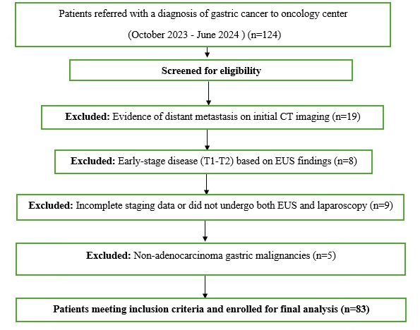
Fig. 1. Flowchart of Patient Selection for the Study
Table 2. Diagnostic Performance of EUS for Each Stage Compared to Laparoscopic Staging
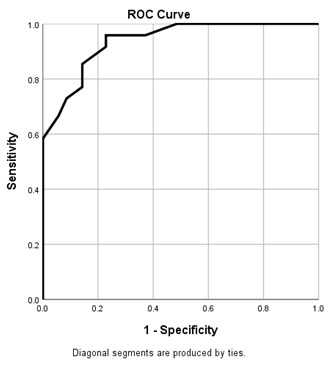
Fig. 2. Receiver Operating Characteristic (ROC) Curve of Tumor Size for Predicting Advanced Gastric Cancer Stage Detected by Laparoscopy. AUC was 0.938 (95% CI: 0.890–0.985, p < 0.001).
Table 3. Contingency Table of EUS versus Laparoscopic Staging
Table 4. Subgroup Analysis of Agreement
Full-Text: (10 Views)
Introduction
Gastric cancer remains a significant global health challenge, ranking as the fifth most common malignancy as well as the third leading cause of cancer-related death worldwide [1]. In Iran, especially in the southeast region, the incidence is notably high, with an age-standardized rate of 22.9 per 100,000 population [2]. Unfortunately, gastric cancer is often diagnosed at advanced stages in this region, which heightens diagnostic and therapeutic complexity and poses a considerable burden on public health systems [3, 4]. In such contexts, timely and accurate staging of the disease is essential for both guiding treatment decisions and improving patient outcomes.
Endoscopic ultrasound (EUS) and staging laparoscopy are two essential diagnostic modalities utilized in the clinical evaluation of gastric cancer. These tools play a key role in determining tumor depth (T), lymph node involvement (N), and the presence of occult peritoneal metastases. Accurate staging allows for better selection of therapeutic strategies, helps prevent unnecessary surgeries, and avoids ineffective or harmful treatments. In low- and middle-income countries (LMICs), where healthcare resources are limited, optimizing diagnostic performance is even more critical to ensure both clinical efficacy and cost-effectiveness [5].
Several international studies have tested the diagnostic value of EUS and laparoscopy either individually or in combination [6, 7]. For instance, Sacerdotianu et al. (2022) reported EUS overall accuracy for T staging 58.53%, with the highest sensitivity reached for the T4 stage, 95.83%, while laparoscopy presented occult peritoneal metastases in 10% of cases [8, 9]. Mocellin et al. (2015) found that combining the two modalities boosted overall diagnostic accuracy to more than 90% [10]. Likewise, a systematic review and meta-analysis demonstrated that staging laparoscopy had an overall sensitivity of 84.6% and specificity of 100% for detecting peritoneal metastases in gastric cancer [11]. In spite of these findings, the routine use of laparoscopy remains debated in some clinical settings, particularly due to logistical and financial barriers [12].
In an Iranian study, endoscopic ultrasonography demonstrated an overall accuracy of 67.9% for T staging (depth of tumor invasion) and 75.4% for N staging (nodal involvement) in patients with gastric cancer, while staging laparoscopy remains valuable for detecting occult peritoneal metastases missed by conventional imaging modalities [13]. Nevertheless, there is still a paucity of data appraising the diagnostic accuracy of these two modalities in combination, especially in real-world settings of LMICs [11, 14]. Important knowledge gaps remain: few studies directly compare EUS and laparoscopy in unselected clinical populations, explore test performance by tumor characteristics, or apply robust statistical approaches such as ROC curve analysis. Further, guidance is lacking regarding the preferred initial staging method in resource-limited, high-volume referral centers where most patients present with advanced diseases [6-13].
Considering these research gaps—particularly the limited data on diagnostic accuracy comparisons between EUS and laparoscopy in real-world settings of low-resource countries—this study aimed to ascertain the diagnostic performance and agreement between endoscopic ultrasound (EUS) and staging laparoscopy in patients with locally advanced gastric cancer. Laparoscopy was considered the reference standard owing to its higher sensitivity in detecting peritoneal metastases. The findings are expected to inform clinical decision-making and optimize staging strategies in high-burden, resource-limited environments.
Materials and Methods
This diagnostic accuracy and agreement study was performed at Afzalipour and Shahid Bahonar Hospitals, two tertiary referral centers affiliated with Kerman University of Medical Sciences in southeast Iran. These public hospitals provide specialized diagnostic and therapeutic services for gastrointestinal malignancies and serve a wide catchment area, including the provinces of Kerman, Sistan and Baluchestan, Hormozgan, and South Khorasan. Both centers are equipped with advanced imaging modalities and staffed by experienced gastrointestinal specialists, making them well-suited for conducting diagnostic test evaluations.
A total of 124 patients with histologically confirmed gastric adenocarcinoma were initially evaluated between October 2023 and June 2024. Of these, 41 patients were excluded based on the following criteria: presence of distant metastases on initial CT imaging (n = 19), early-stage disease (T1–T2) identified on EUS (n = 8), incomplete staging data or absence of either EUS or laparoscopy (n = 9), and non-adenocarcinoma histology (n = 5). Hence, 83 patients fulfilled the final inclusion criteria and were analyzed. Fig. 1 presents the flowchart of patient selection, including inclusion and exclusion criteria.
This research was a retrospective diagnostic accuracy study; however, a post-hoc power calculation using STATA confirmed that the sample size (n = 83) provided over 80% power to detect statistically significant differences in diagnostic performance between EUS and laparoscopy, assuming a medium effect size and α = 0.05 (two-tailed).
Diagnostic Procedures
Endoscopic Ultrasound (EUS): All EUS procedures were performed using a linear echoendoscope (e.g., Olympus GF-UCT180 or equivalent) under conscious sedation. Experienced gastroenterologists (each with over 5 years of post-fellowship experience in interventional EUS) performed the examinations. The procedure involved a systematic assessment of the gastric wall layers to determine the depth of tumor invasion (T stage) as well as identification of perigastric and celiac axis lymph nodes for assessing nodal involvement (N stage). Criteria for malignant lymph nodes included round shape, hypoechoic texture, sharp borders, and size >1 cm. The entire staging was documented according to the 8th edition of the American Joint Committee on Cancer (AJCC) TNM classification system [15].
Staging Laparoscopy: Staging laparoscopy was performed in the operating room under general anesthesia by fellowship-trained surgical oncologists using a standardized protocol. Following insufflation and placement of trocars (typically via three-port access), a systematic exploration of the peritoneal cavity, anterior and posterior liver surfaces, diaphragm, lesser sac, omentum, and pelvis was conducted. Peritoneal lavage was conducted for cytology. Any suspicious lesions (nodules, discolorations, thickening) were biopsied and sent for frozen section or histopathologic confirmation [16]. The findings were documented and compared with those from EUS. If distant metastases (M1 disease) were confirmed, the patient was referred for non-surgical management [17].
TNM Classification: Tumors were staged according to the TNM classification system. For T staging, tumors were categorized based on the depth of invasion as follows: T1a indicated invasion limited to the lamina propria or muscularis mucosae; T1b involved the submucosa; T2 extended into the muscularis propria; T3 reached the subserosal layer; and T4 denoted invasion into the serosa (T4a) or adjacent structures (T4b) [15].
For N staging, nodal involvement was classified as N0 when no regional lymph node metastases were observed, N1 for 1–2 positive nodes, N2 for 3–6 nodes, N3a for 7–15 nodes, and N3b when more than 15 regional lymph nodes were involved. M staging is distinguished between M0 (no evidence of distant metastasis) and M1 (presence of distant metastasis) [17].
We analyzed data using SPSS v26 and STATA v17. Cohen’s kappa was utilized to assess agreement between EUS and laparoscopy, supplemented by diagnostic accuracy metrics and McNemar’s test to compare discordant cases. Sensitivity, specificity, positive predictive value (PPV), negative predictive value (NPV), overall accuracy, and the area under the receiver operating characteristic curve (AUC) were calculated, where laparoscopy served as the reference standard. A power calculation was carried out to justify the sample size (n = 83), confirming 80% power to detect a statistically significant difference in diagnostic accuracy (alpha = 0.05, two-tailed), assuming a moderate effect size [10]. Subgroup analyses were undertaken to ascertain diagnostic agreement across tumor size, location, and H. pylori status (Table 4). A p-value < 0.05 was considered statistically significant for all tests.
Gastric cancer remains a significant global health challenge, ranking as the fifth most common malignancy as well as the third leading cause of cancer-related death worldwide [1]. In Iran, especially in the southeast region, the incidence is notably high, with an age-standardized rate of 22.9 per 100,000 population [2]. Unfortunately, gastric cancer is often diagnosed at advanced stages in this region, which heightens diagnostic and therapeutic complexity and poses a considerable burden on public health systems [3, 4]. In such contexts, timely and accurate staging of the disease is essential for both guiding treatment decisions and improving patient outcomes.
Endoscopic ultrasound (EUS) and staging laparoscopy are two essential diagnostic modalities utilized in the clinical evaluation of gastric cancer. These tools play a key role in determining tumor depth (T), lymph node involvement (N), and the presence of occult peritoneal metastases. Accurate staging allows for better selection of therapeutic strategies, helps prevent unnecessary surgeries, and avoids ineffective or harmful treatments. In low- and middle-income countries (LMICs), where healthcare resources are limited, optimizing diagnostic performance is even more critical to ensure both clinical efficacy and cost-effectiveness [5].
Several international studies have tested the diagnostic value of EUS and laparoscopy either individually or in combination [6, 7]. For instance, Sacerdotianu et al. (2022) reported EUS overall accuracy for T staging 58.53%, with the highest sensitivity reached for the T4 stage, 95.83%, while laparoscopy presented occult peritoneal metastases in 10% of cases [8, 9]. Mocellin et al. (2015) found that combining the two modalities boosted overall diagnostic accuracy to more than 90% [10]. Likewise, a systematic review and meta-analysis demonstrated that staging laparoscopy had an overall sensitivity of 84.6% and specificity of 100% for detecting peritoneal metastases in gastric cancer [11]. In spite of these findings, the routine use of laparoscopy remains debated in some clinical settings, particularly due to logistical and financial barriers [12].
In an Iranian study, endoscopic ultrasonography demonstrated an overall accuracy of 67.9% for T staging (depth of tumor invasion) and 75.4% for N staging (nodal involvement) in patients with gastric cancer, while staging laparoscopy remains valuable for detecting occult peritoneal metastases missed by conventional imaging modalities [13]. Nevertheless, there is still a paucity of data appraising the diagnostic accuracy of these two modalities in combination, especially in real-world settings of LMICs [11, 14]. Important knowledge gaps remain: few studies directly compare EUS and laparoscopy in unselected clinical populations, explore test performance by tumor characteristics, or apply robust statistical approaches such as ROC curve analysis. Further, guidance is lacking regarding the preferred initial staging method in resource-limited, high-volume referral centers where most patients present with advanced diseases [6-13].
Considering these research gaps—particularly the limited data on diagnostic accuracy comparisons between EUS and laparoscopy in real-world settings of low-resource countries—this study aimed to ascertain the diagnostic performance and agreement between endoscopic ultrasound (EUS) and staging laparoscopy in patients with locally advanced gastric cancer. Laparoscopy was considered the reference standard owing to its higher sensitivity in detecting peritoneal metastases. The findings are expected to inform clinical decision-making and optimize staging strategies in high-burden, resource-limited environments.
Materials and Methods
This diagnostic accuracy and agreement study was performed at Afzalipour and Shahid Bahonar Hospitals, two tertiary referral centers affiliated with Kerman University of Medical Sciences in southeast Iran. These public hospitals provide specialized diagnostic and therapeutic services for gastrointestinal malignancies and serve a wide catchment area, including the provinces of Kerman, Sistan and Baluchestan, Hormozgan, and South Khorasan. Both centers are equipped with advanced imaging modalities and staffed by experienced gastrointestinal specialists, making them well-suited for conducting diagnostic test evaluations.
A total of 124 patients with histologically confirmed gastric adenocarcinoma were initially evaluated between October 2023 and June 2024. Of these, 41 patients were excluded based on the following criteria: presence of distant metastases on initial CT imaging (n = 19), early-stage disease (T1–T2) identified on EUS (n = 8), incomplete staging data or absence of either EUS or laparoscopy (n = 9), and non-adenocarcinoma histology (n = 5). Hence, 83 patients fulfilled the final inclusion criteria and were analyzed. Fig. 1 presents the flowchart of patient selection, including inclusion and exclusion criteria.
This research was a retrospective diagnostic accuracy study; however, a post-hoc power calculation using STATA confirmed that the sample size (n = 83) provided over 80% power to detect statistically significant differences in diagnostic performance between EUS and laparoscopy, assuming a medium effect size and α = 0.05 (two-tailed).
Diagnostic Procedures
Endoscopic Ultrasound (EUS): All EUS procedures were performed using a linear echoendoscope (e.g., Olympus GF-UCT180 or equivalent) under conscious sedation. Experienced gastroenterologists (each with over 5 years of post-fellowship experience in interventional EUS) performed the examinations. The procedure involved a systematic assessment of the gastric wall layers to determine the depth of tumor invasion (T stage) as well as identification of perigastric and celiac axis lymph nodes for assessing nodal involvement (N stage). Criteria for malignant lymph nodes included round shape, hypoechoic texture, sharp borders, and size >1 cm. The entire staging was documented according to the 8th edition of the American Joint Committee on Cancer (AJCC) TNM classification system [15].
Staging Laparoscopy: Staging laparoscopy was performed in the operating room under general anesthesia by fellowship-trained surgical oncologists using a standardized protocol. Following insufflation and placement of trocars (typically via three-port access), a systematic exploration of the peritoneal cavity, anterior and posterior liver surfaces, diaphragm, lesser sac, omentum, and pelvis was conducted. Peritoneal lavage was conducted for cytology. Any suspicious lesions (nodules, discolorations, thickening) were biopsied and sent for frozen section or histopathologic confirmation [16]. The findings were documented and compared with those from EUS. If distant metastases (M1 disease) were confirmed, the patient was referred for non-surgical management [17].
TNM Classification: Tumors were staged according to the TNM classification system. For T staging, tumors were categorized based on the depth of invasion as follows: T1a indicated invasion limited to the lamina propria or muscularis mucosae; T1b involved the submucosa; T2 extended into the muscularis propria; T3 reached the subserosal layer; and T4 denoted invasion into the serosa (T4a) or adjacent structures (T4b) [15].
For N staging, nodal involvement was classified as N0 when no regional lymph node metastases were observed, N1 for 1–2 positive nodes, N2 for 3–6 nodes, N3a for 7–15 nodes, and N3b when more than 15 regional lymph nodes were involved. M staging is distinguished between M0 (no evidence of distant metastasis) and M1 (presence of distant metastasis) [17].
We analyzed data using SPSS v26 and STATA v17. Cohen’s kappa was utilized to assess agreement between EUS and laparoscopy, supplemented by diagnostic accuracy metrics and McNemar’s test to compare discordant cases. Sensitivity, specificity, positive predictive value (PPV), negative predictive value (NPV), overall accuracy, and the area under the receiver operating characteristic curve (AUC) were calculated, where laparoscopy served as the reference standard. A power calculation was carried out to justify the sample size (n = 83), confirming 80% power to detect a statistically significant difference in diagnostic accuracy (alpha = 0.05, two-tailed), assuming a moderate effect size [10]. Subgroup analyses were undertaken to ascertain diagnostic agreement across tumor size, location, and H. pylori status (Table 4). A p-value < 0.05 was considered statistically significant for all tests.
Fig. 1. Flowchart of Patient Selection for the Study
Results
A total of 83 patients were included in the study, with a mean age of 64.99 ± 9.84 years (range: 36–82), and 61.4% of the participants were male. The risk factor
profile of the study population presented a history of smoking in 22.9% of patients, previous endoscopy in 16.9%, and Helicobacter pylori infection in 12.0%. None of the patients reported alcohol consumption (Table 1).
A total of 83 patients were included in the study, with a mean age of 64.99 ± 9.84 years (range: 36–82), and 61.4% of the participants were male. The risk factor
profile of the study population presented a history of smoking in 22.9% of patients, previous endoscopy in 16.9%, and Helicobacter pylori infection in 12.0%. None of the patients reported alcohol consumption (Table 1).
Table 1. Demographic Characteristics and Disease Stage Distribution Based on EUS and Laparoscopy in Patients with Gastric Cancer (n = 83)
| Characteristic | Value / Frequency (%) | |
| Age (years), mean ± SD | 64.99 ± 9.84 | |
| Age range (years) | 36–82 | |
| Gender | Male | 51 (61.4) |
| Female | 32 (38.6) | |
| Risk factors | Smoking history | 19 (22.9) |
| Family history | 5 (6.0) | |
| Helicobacter pylori infection | 10 (12.0) | |
| Previous endoscopy | 14 (16.9) | |
| Alcohol consumption | 0 (0) | |
| EUS TNM Staging | T3 | 51 (61.4) |
| T4 | 32 (38.6) | |
| N1 | 58 (69.9) | |
| N2 | 25 (30.1) | |
| M0 | 83 (100) | |
| M1 | 0 (0) | |
| Overall Stage by EUS | Stage I | 0 (0) |
| Stage II | 39 (47.0) | |
| Stage III | 44 (53.0) | |
| Stage IV | 0 (0) | |
| Overall Stage by Laparoscopy | Stage I | 0 (0) |
| Stage II | 35 (42.2) | |
| Stage III | 47 (56.6) | |
| Stage IV | 1 (1.2) | |
According to endoscopic ultrasound (EUS) assessment, 61.4% of patients were staged as T3 and 38.6% as T4. Nodal involvement was reported as N1 in 69.9% and N2 in 30.1%, whereas all patients were classified as M0 based on EUS. Regarding TNM stage grouping by EUS, no patients were classified as stage I or IV. Instead, 47.0% of patients were placed in stage II and 53.0% in stage III. In comparison, laparoscopic staging categorized 42.2% of patients as stage II, 56.6% as stage III, and 1.2% (n = 1) as stage IV (Table 1).
In order to ascertain the diagnostic accuracy of EUS, laparoscopic staging was considered the reference standard. When distinguishing advanced-stage disease (Stage III or IV) from early-stage disease (Stage II), EUS revealed a sensitivity of 91.7%, specificity of 100%, positive predictive value (PPV) of 100%, and negative predictive value (NPV) of 89.7% (Table 2). These values indicate that EUS has high diagnostic performance in distinguishing advanced gastric cancer from non-advanced cases.
In order to ascertain the diagnostic accuracy of EUS, laparoscopic staging was considered the reference standard. When distinguishing advanced-stage disease (Stage III or IV) from early-stage disease (Stage II), EUS revealed a sensitivity of 91.7%, specificity of 100%, positive predictive value (PPV) of 100%, and negative predictive value (NPV) of 89.7% (Table 2). These values indicate that EUS has high diagnostic performance in distinguishing advanced gastric cancer from non-advanced cases.
Table 2. Diagnostic Performance of EUS for Each Stage Compared to Laparoscopic Staging
| Stage | TP | FP | FN | TN | Sensitivity (%) | Specificity (%) | PPV (%) | NPV (%) | Cohen's Kappa | 95% CI (for Kappa or Metric), (P-value) |
| II | 35 | 0 | 4 | 44 | 89.7 | 100 | 100 | 91.7 | — | — |
| III-IV | 44 | 0 | 4 | 35 | 91.7 | 100 | 100 | 89.7 |
| Overall | — | — | — | — | — | — | — | — | 0.88 | )0.77–0.98(, )p < 0.001( |
- TP = True Positive: Cases correctly classified as advanced stage (III–IV)
- FP = False Positive: Cases incorrectly classified as advanced by EUS
- TN = True Negative: Cases correctly classified as early stage (I–II)
- FN = False Negative: Advanced cases missed by EUS
- Statistical tests used: Sensitivity, specificity, PPV, and NPV were calculated using standard diagnostic test formulas. Kappa statistics were used to assess agreement between EUS and laparoscopic staging. Confidence intervals were calculated using the Wilson method.
Fig. 2. Receiver Operating Characteristic (ROC) Curve of Tumor Size for Predicting Advanced Gastric Cancer Stage Detected by Laparoscopy. AUC was 0.938 (95% CI: 0.890–0.985, p < 0.001).
Receiver operating characteristic (ROC) curve analysis was performed to evaluate the ability of tumor size to predict advanced-stage gastric cancer (Stage III–IV versus Stage II) based on laparoscopic staging. The area under the ROC curve (AUC) was 0.938 (95% CI: 0.890–0.985, p < 0.001), demonstrating excellent discriminative ability (Fig. 2). These findings indicate that increasing tumor size is strongly associated with advanced-stage disease detected during staging
laparoscopy.
Table 3 presents the cross-tabulation of staging results obtained by endoscopic ultrasound (EUS) and staging laparoscopy. Overall, a high level of agreement was observed between the two modalities in distinguishing Stage II from Stage III/IV disease. Most patients were consistently classified across both methods, with only a small number of discordant cases identified during laparoscopic assessment.
laparoscopy.
Table 3 presents the cross-tabulation of staging results obtained by endoscopic ultrasound (EUS) and staging laparoscopy. Overall, a high level of agreement was observed between the two modalities in distinguishing Stage II from Stage III/IV disease. Most patients were consistently classified across both methods, with only a small number of discordant cases identified during laparoscopic assessment.
Table 3. Contingency Table of EUS versus Laparoscopic Staging
| Total | Laparoscopic stage | EUS stage | ||
| IV | III | II | ||
| 39 | 0 | 4 | 35 | II |
| 44 | 1 | 43 | 0 | III |
| 83 | 1 | 47 | 35 | Total |
| Cohen's kappa coefficient: 0.88 (95% CI: 0.77-0.98, p<0.001) | ||||
Statistical tests used: Cohen’s kappa coefficient was computed to assess the level of agreement between staging modalities. P-value derived from test of agreement (two-tailed).
The overall agreement between EUS and laparoscopy in TNM staging (Stage II vs. III/IV) was 94.0%. Cohen’s kappa coefficient was 0.88 (95% CI: 0.77–0.98, p < 0.001), reflecting near-perfect agreement between the two modalities.
Subgroup analyses further supported the robustness of this agreement across different demographic and tumor-related factors (Table 4). The largest kappa values were observed among patients with tumor size ≥5 cm (kappa = 0.91), those with distal or mid-gastric tumors (kappa = 0.90), and H. pylori-positive individuals (kappa = 0.95). Agreement was also consistent among males (kappa = 0.89), females (kappa = 0.88), patients aged <65 years (kappa = 0.87), and those with a history of smoking (kappa = 0.85). Laparoscopy led to upstaging in five patients (6.0% of cases). Four patients were upstaged from Stage II to III based on peritoneal seeding identified in the omentum and pelvic peritoneum, whereas one patient was upstaged to Stage IV because of biopsy-confirmed liver metastases. These findings highlight the added clinical value of staging laparoscopy in identifying peritoneal or hepatic involvement that may not be visible on EUS, and which could alter clinical management from curative surgery to neoadjuvant chemotherapy or palliative approaches.
Nevertheless, since no pathological verification was available for nodal staging, the accuracy metrics for N staging could not be calculated. Future studies with surgical or histopathologic follow-up are required to validate EUS findings in nodal assessment.
Subgroup analyses further supported the robustness of this agreement across different demographic and tumor-related factors (Table 4). The largest kappa values were observed among patients with tumor size ≥5 cm (kappa = 0.91), those with distal or mid-gastric tumors (kappa = 0.90), and H. pylori-positive individuals (kappa = 0.95). Agreement was also consistent among males (kappa = 0.89), females (kappa = 0.88), patients aged <65 years (kappa = 0.87), and those with a history of smoking (kappa = 0.85). Laparoscopy led to upstaging in five patients (6.0% of cases). Four patients were upstaged from Stage II to III based on peritoneal seeding identified in the omentum and pelvic peritoneum, whereas one patient was upstaged to Stage IV because of biopsy-confirmed liver metastases. These findings highlight the added clinical value of staging laparoscopy in identifying peritoneal or hepatic involvement that may not be visible on EUS, and which could alter clinical management from curative surgery to neoadjuvant chemotherapy or palliative approaches.
Nevertheless, since no pathological verification was available for nodal staging, the accuracy metrics for N staging could not be calculated. Future studies with surgical or histopathologic follow-up are required to validate EUS findings in nodal assessment.
Table 4. Subgroup Analysis of Agreement
| Subgroup | N | Kappa | 95% CI |
| Tumor size <5 cm | 29 | 0.83 | 0.68-0.98 |
| Tumor size ≥5 cm | 54 | 0.91 | 0.82-1.00 |
| Distal/mid-gastric | 62 | 0.90 | 0.80-1.00 |
| Proximal | 21 | 0.84 | 0.67-1.00 |
| H. pylori positive | 10 | 0.95 | 0.85-1.00 |
| Age <65 years | 45 | 0.87 | 0.75-0.99 |
| Age ≥65 years | 38 | 0.89 | 0.78-1.00 |
| Male | 51 | 0.89 | 0.78-1.00 |
| Female | 32 | 0.88 | 0.76-1.00 |
| Smoking history | 19 | 0.85 | 0.70-1.00 |
Statistical tests used: Subgroup-specific Cohen’s kappa values and 95% confidence intervals were calculated using the Fleiss-Cohen method. No formal statistical test for subgroup comparison was applied due to sample size limitations.
Discussion
This study appraised the diagnostic accuracy and staging agreement between endoscopic ultrasound (EUS) and staging laparoscopy in patients with gastric cancer in southeast Iran. Our findings revealed a strong agreement (κ = 0.88, 95% CI: 0.77–0.98, p < 0.001) between the two modalities in distinguishing stage II versus stage III/IV disease, signaling a high level of concordance. Nevertheless, staging laparoscopy led to upstaging 6% of patients owing to the detection of peritoneal or liver metastases that were missed in EUS. This underscores the clinical value of laparoscopy in revealing occult advanced disease that may alter therapeutic plans.
Several previous studies support our findings, considering the limitations of EUS in detecting peritoneal dissemination. For instance, Levy et al. (2015) and Lee et al. (2005) reported that while EUS is useful for ascertaining T and N stages, it often fails to identify serosal invasion or peritoneal metastasis [18, 19]. Likewise, Van Hootegem et al. (2025) emphasized that staging laparoscopy has superior sensitivity for detecting small-volume peritoneal spread, which can be missed by imaging alone [20]. These studies concur with our finding that EUS, though valuable for locoregional staging, is insufficient as a standalone tool for comprehensive staging in advanced gastric cancer.
A meta-analysis by Puli et al. (2008) also reported high diagnostic performance of EUS in appraising T3/T4 tumors but noted lowered accuracy when distant metastasis is present [17]. Further, Shi et al. (2019) highlighted that EUS accuracy is affected by operator expertise, tumor location, as well as the presence of ascites or serosal involvement—all potential sources of variability in real-world settings like ours [21].
In contrast, some studies, such as those by Mocellin et al. (2011) and Nucci et al. (2023), documented higher staging concordance (κ > 0.90) between EUS and surgical or laparoscopic staging. These differences may emanate from more selective inclusion criteria or exclusion of patients with advanced or metastatic disease, thus artificially enhancing EUS performance in earlier-stage cancers [4, 22]. In contrast, our study reflects a real-world clinical population with mostly locally advanced disease, which heightens the staging challenge.
A unique finding in our study was the low observed prevalence of Helicobacter pylori infection (12%). This is notably lower than previous national studies in Iran, which reported H. pylori prevalence rates of 60–80% in adults [23, 24]. Possible explanations include undocumented prior antibiotic use, histologic masking of infection in late-stage cancers, or underreporting in clinical records. This discrepancy highlights the need for more systematic documentation of infection status in oncology registries.
Taken together, these findings support the usage of a complementary staging strategy using both EUS and laparoscopy [25]. While EUS remains essential for ascertaining locoregional invasion and lymphadenopathy, it should not be solely relied upon to rule out peritoneal metastases. Diagnostic laparoscopy remains critical, especially in patients who are surgical candidates or are being considered for neoadjuvant chemotherapy.
This study offered several notable strengths. By focusing on a region with limited data on gastric cancer staging [26], it addressed a critical gap in the literature. The robust methodology, including the use of laparoscopy as the reference standard and comprehensive statistical analyses (e.g., Cohen’s kappa and ROC curves), could enhance the reliability of the findings. Further, subgroup analyses based on tumor size, location, and Helicobacter pylori status would provide nuanced insights that can guide personalized clinical decision-making.
Nevertheless, certain limitations should be acknowledged to fully interpret the results. As the study was performed at two tertiary centers in a single geographic region, its findings may not be fully generalizable to other populations or healthcare settings. Further, the absence of long-term follow-up data limits our ability to appraise the impact of staging modifications on patient outcomes. In order to address these constraints, future prospective multicenter studies with larger and more diverse cohorts could validate and extend our findings. Further investigation of the cost-effectiveness of routine staging laparoscopy in specific patient subgroups could optimize resource allocation. Eventually, exploring the integration of emerging molecular and imaging biomarkers with conventional staging modalities could further refine diagnostic algorithms, potentially ameliorating accuracy and patient outcomes.
Conclusion
This study demonstrated a strong diagnostic agreement between endoscopic ultrasound (EUS) and staging laparoscopy for TNM staging (Stage II vs. III/IV), with 94.0% concordance (κ = 0.88, 95% CI: 0.77–0.98, p < 0.001). Laparoscopy upstaged 6% of cases by detecting peritoneal or liver metastases missed by EUS, often in T3 or higher stages, underscoring its role in identifying occult advanced disease that may alter treatment. Subgroup analyses revealed a robust agreement, particularly for tumors ≥5 cm (κ = 0.91) and H. pylori-positive cases (κ = 0.95). The ROC curve (AUC = 0.938) confirmed EUS’s high discriminative ability. A targeted combined approach could boost accuracy and cost-effectiveness in resource-limited settings such as southeastern Iran.
Acknowledgements
We thank the Clinical Research Development Unit of Bahonar Hospital, Kerman University of Medical Sciences, for their support.
Conflict of interest
None declared.
Funding
This study was supported by the Kerman University of Medical Sciences.
Ethical Considerations
The study was conducted in accordance with ethical principles for diagnostic test evaluation studies.
Code of Ethics
The study protocol was approved by the Ethics Committee of Kerman University of Medical Sciences (Ethics Code: IR.KMU.AH.REC.1403.107)
Authors' Contributions
Mohammad Karim Pouramiri: Conceptualization; Methodology; Investigation; Resources; Writing Original Draft; Project administration; Mohammad
Shafiei: Conceptualization; Methodology; Validation; Formal analysis; Investigation; Writing – Review & Editing; Visualization; Fatemeh Doost-Mohammadi: Methodology; Software; Formal analysis; Data Curation; Writing – Review & Editing; Supervision (epidemiological/statistical aspects); Alireza Amirbeigi: Conceptualization; Methodology; Validation; Investigation; Resources; Writing – Review & Editing; Supervision; Funding acquisition; Correspondence.
This study appraised the diagnostic accuracy and staging agreement between endoscopic ultrasound (EUS) and staging laparoscopy in patients with gastric cancer in southeast Iran. Our findings revealed a strong agreement (κ = 0.88, 95% CI: 0.77–0.98, p < 0.001) between the two modalities in distinguishing stage II versus stage III/IV disease, signaling a high level of concordance. Nevertheless, staging laparoscopy led to upstaging 6% of patients owing to the detection of peritoneal or liver metastases that were missed in EUS. This underscores the clinical value of laparoscopy in revealing occult advanced disease that may alter therapeutic plans.
Several previous studies support our findings, considering the limitations of EUS in detecting peritoneal dissemination. For instance, Levy et al. (2015) and Lee et al. (2005) reported that while EUS is useful for ascertaining T and N stages, it often fails to identify serosal invasion or peritoneal metastasis [18, 19]. Likewise, Van Hootegem et al. (2025) emphasized that staging laparoscopy has superior sensitivity for detecting small-volume peritoneal spread, which can be missed by imaging alone [20]. These studies concur with our finding that EUS, though valuable for locoregional staging, is insufficient as a standalone tool for comprehensive staging in advanced gastric cancer.
A meta-analysis by Puli et al. (2008) also reported high diagnostic performance of EUS in appraising T3/T4 tumors but noted lowered accuracy when distant metastasis is present [17]. Further, Shi et al. (2019) highlighted that EUS accuracy is affected by operator expertise, tumor location, as well as the presence of ascites or serosal involvement—all potential sources of variability in real-world settings like ours [21].
In contrast, some studies, such as those by Mocellin et al. (2011) and Nucci et al. (2023), documented higher staging concordance (κ > 0.90) between EUS and surgical or laparoscopic staging. These differences may emanate from more selective inclusion criteria or exclusion of patients with advanced or metastatic disease, thus artificially enhancing EUS performance in earlier-stage cancers [4, 22]. In contrast, our study reflects a real-world clinical population with mostly locally advanced disease, which heightens the staging challenge.
A unique finding in our study was the low observed prevalence of Helicobacter pylori infection (12%). This is notably lower than previous national studies in Iran, which reported H. pylori prevalence rates of 60–80% in adults [23, 24]. Possible explanations include undocumented prior antibiotic use, histologic masking of infection in late-stage cancers, or underreporting in clinical records. This discrepancy highlights the need for more systematic documentation of infection status in oncology registries.
Taken together, these findings support the usage of a complementary staging strategy using both EUS and laparoscopy [25]. While EUS remains essential for ascertaining locoregional invasion and lymphadenopathy, it should not be solely relied upon to rule out peritoneal metastases. Diagnostic laparoscopy remains critical, especially in patients who are surgical candidates or are being considered for neoadjuvant chemotherapy.
This study offered several notable strengths. By focusing on a region with limited data on gastric cancer staging [26], it addressed a critical gap in the literature. The robust methodology, including the use of laparoscopy as the reference standard and comprehensive statistical analyses (e.g., Cohen’s kappa and ROC curves), could enhance the reliability of the findings. Further, subgroup analyses based on tumor size, location, and Helicobacter pylori status would provide nuanced insights that can guide personalized clinical decision-making.
Nevertheless, certain limitations should be acknowledged to fully interpret the results. As the study was performed at two tertiary centers in a single geographic region, its findings may not be fully generalizable to other populations or healthcare settings. Further, the absence of long-term follow-up data limits our ability to appraise the impact of staging modifications on patient outcomes. In order to address these constraints, future prospective multicenter studies with larger and more diverse cohorts could validate and extend our findings. Further investigation of the cost-effectiveness of routine staging laparoscopy in specific patient subgroups could optimize resource allocation. Eventually, exploring the integration of emerging molecular and imaging biomarkers with conventional staging modalities could further refine diagnostic algorithms, potentially ameliorating accuracy and patient outcomes.
Conclusion
This study demonstrated a strong diagnostic agreement between endoscopic ultrasound (EUS) and staging laparoscopy for TNM staging (Stage II vs. III/IV), with 94.0% concordance (κ = 0.88, 95% CI: 0.77–0.98, p < 0.001). Laparoscopy upstaged 6% of cases by detecting peritoneal or liver metastases missed by EUS, often in T3 or higher stages, underscoring its role in identifying occult advanced disease that may alter treatment. Subgroup analyses revealed a robust agreement, particularly for tumors ≥5 cm (κ = 0.91) and H. pylori-positive cases (κ = 0.95). The ROC curve (AUC = 0.938) confirmed EUS’s high discriminative ability. A targeted combined approach could boost accuracy and cost-effectiveness in resource-limited settings such as southeastern Iran.
Acknowledgements
We thank the Clinical Research Development Unit of Bahonar Hospital, Kerman University of Medical Sciences, for their support.
Conflict of interest
None declared.
Funding
This study was supported by the Kerman University of Medical Sciences.
Ethical Considerations
The study was conducted in accordance with ethical principles for diagnostic test evaluation studies.
Code of Ethics
The study protocol was approved by the Ethics Committee of Kerman University of Medical Sciences (Ethics Code: IR.KMU.AH.REC.1403.107)
Authors' Contributions
Mohammad Karim Pouramiri: Conceptualization; Methodology; Investigation; Resources; Writing Original Draft; Project administration; Mohammad
Shafiei: Conceptualization; Methodology; Validation; Formal analysis; Investigation; Writing – Review & Editing; Visualization; Fatemeh Doost-Mohammadi: Methodology; Software; Formal analysis; Data Curation; Writing – Review & Editing; Supervision (epidemiological/statistical aspects); Alireza Amirbeigi: Conceptualization; Methodology; Validation; Investigation; Resources; Writing – Review & Editing; Supervision; Funding acquisition; Correspondence.
References
1. Bray F, Laversanne M, Sung H, Ferlay J, Siegel RL, Soerjomataram I, et al. Global Cancer Statistics 2022: GLOBOCAN estimates of incidence and mortality worldwide for 36 cancers in 185 countries. CA Cancer J Clin. 2024;74(3):229-63. [DOI]
2. Fattahi N, Moghaddam SS, Rezaei N, Rezaei N, Fattahi E, Moradveisi B, et al. The national trend of the gastric cancer burden in Iran from 1990-2017. Asia Pac J Clin Oncol. 2022;18(2):e96-102. [DOI] [PMID]
3. Zhu Z, Gong Y, Xu H. Clinical and pathological staging of gastric cancer: Current perspectives and implications. Eur J Surg Oncol. 2020;46(10 Pt B):e14-e9. [DOI] [PMID]
4. Mocellin S, Marchet A, Nitti D. EUS for the staging of gastric cancer: a meta-analysis. Gastrointest Endosc. 2011;73(6):1122-34. [DOI] [PMID]
5. Sacerdotianu VM, Ungureanu BS, Iordache S, Filip MM, Pirici D, Liliac IM, et al. Accuracy of endoscopic ultrasonography for gastric cancer staging. Curr Health Sci J. 2022;48(1):88-94. [DOI] [PMID] [PMCID]
6. Schenck R, Sellers E, Walsh L, Zaydfudim VM, Bauer TW, Feng Su, et al. Utility of Endoscopic Ultrasound in Gastric Cancer Staging. Gastrointest Endosc. 2022; 95(6):AB486. [DOI]
7. Ziogas DI, Kalakos N, Manolakis A, Voulgaris T, Vezakis I, Tadic M, et al. Endoscopic ultrasound (EUS) in gastric cancer: current applications and future perspectives. Diseases. 2025;13(8):234. [DOI]
8. Sacerdotianu VM, Ungureanu BS, Iordache S, Filip MM, Pirici D, Liliac IM, et al. Accuracy of Endoscopic Ultrasonography for Gastric Cancer Staging. Curr Health Sci J. 2022;48(1):88-94. [DOI] [PMID] [PMCID]
9. Sugarbaker PH. Laparoscopy in the diagnosis and treatment of peritoneal metastases. Ann Laparosc Endosc Surg. 2019;4. [URL]
10. Mocellin S, Pasquali S. Diagnostic accuracy of endoscopic ultrasonography (EUS) for the preoperative locoregional staging of primary gastric cancer. Cochrane Database of Syst Rev. 2015;2. [DOI]
11. Ramos RF, Scalon FM, Scalon MM, Dias DI. Staging laparoscopy in gastric cancer to detect peritoneal metastases: A systematic review and meta-analysis. Eur J Surg Oncol. 2016;42(9):1315-21. [DOI]
12. Li K, Cannon JGD, Jiang SY, Sambare TD, Owens DK, Bendavid E, et al. Diagnostic staging laparoscopy in gastric cancer treatment: A cost-effectiveness analysis. J Surg Oncol. 2018;117(6):1288-96. [DOI] [PMID]
13. Razavi SM, Khodadost M, Sohrabi M, Keshavarzi A, Zamani F, Rakhshani N, et al. Accuracy of Endoscopic Ultrasonography for Determination of Tumor Invasion Depth in Gastric Cancer. Asian Pac J Cancer Prev. 2015;16(8):3141-5. [Article]
14. Groh EM, Gupta S, Brown ZJ, Enewold L, Gamble LA, Hernandez JM, et al. Staging Laparoscopy is Underutilized in the Management of Gastric Adenocarcinoma. Ann Surg Oncol. 2020;27(5):1473-9. [DOI] [PMID] [PMCID]
15. Amin MB, Gress DM, Meyer Vega LR, Edge SB, Greene FL, Byrd DR, et al. editors. AJCC Cancer Staging Manual. 8th ed. New York, United States: Springer; 2018. [URL]
16. Hafford ML, Sickle KRV, Willis RE, Wilson TD, Gugliuzza K, Brown KM, et al. Ensuring competency: Are fundamentals of laparoscopic surgery training and certification necessary for practicing surgeons and operating room personnel? Surgical Endoscopy. 2012;27:118-26. [DOI]
17. Puli SR, Reddy JB, Bechtold ML, Antillon D, Ibdah JA, Antillon MR. Staging accuracy of endoscopic ultrasound in gastric carcinoma: meta-analysis and systematic review. World J Gastroenterol. 2008;14(10):1479-90. [DOI] [PMID] [PMCID]
18. Levy MJ, Abu Dayyeh BK, Fujii LL, Clayton AC, Reynolds JP, Lopes TL, et al. Detection of peritoneal carcinomatosis by EUS fine-needle aspiration: impact on staging and resectability (with videos). Gastrointest Endosc. 2015;81(5):1215-24. [DOI]
19. Lee YT, Ng EKW, Hung LCT, Chung SCS, Ching JYL, Chan WY, et al. Accuracy of endoscopic ultrasonography in diagnosing ascites and predicting peritoneal metastases in gastric cancer patients. Gut. 2006;54(11):1524. [DOI]
20. Van Hootegem SJM, Guchelaar NAD, van der Sluis K., Triemstra L, Mönig SP, Rawicz-Pruszyński K, et al. Staging laparoscopy for gastric cancer: European consensus. Br J Surg. 2025; 112(9):znaf144. [DOI] [PMID] [PMCID]
21. Shi D, Xi XX. Factors Affecting the Accuracy of Endoscopic Ultrasonography in the Diagnosis of Early Gastric Cancer Invasion Depth: A Meta-analysis. Gastroenterol Res Pract. 2019;2019(1):8241381. [DOI]
22. de Nucci G, Gabbani T, Impellizzeri G, Deiana S, Biancheri P, Ottaviani L, et al. Linear EUS accuracy in preoperative staging of gastric cancer: a retrospective multicenter study. Diagnostics. 2023;13(11):1842. [DOI]
23. Esmaeilzadeh A, Goshayeshi L, Bergquist R, Jarahi L, Khooei A, Fazeli A, et al. Characteristics of gastric precancerous conditions and Helicobacter pylori infection among dyspeptic patients in north-eastern Iran: Is endoscopic biopsy and histopathological assessment necessary?. BMC Cancer. 2021;21:1143. [DOI]
24. Ashtari S, Pourhoseingholi MA, Molaei M, Taslimi H, Zali MR. The prevalence of Helicobacter pylori is decreasing in Iranian patients. Gastroenterol Hepatol Bed Bench. 2015 Spring;8(Suppl 1):S23-9. [PMID] [PMCID]
25. Power DG, Schattner MA, Gerdes H, Brenner B, Markowitz AJ, Capanu M, et al. Endoscopic Ultrasound Can Improve the Selection for Laparoscopy in Patients with Localized Gastric Cancer. J Am Coll Surg. 2009;208(2):173-8. [DOI]
26. Vazirinejad R, Najafipour R, Rezaeian M, Ghazizadeh A, Doost Mohammadi F. Opium and risk of gastrointestinal cancer: a case-control study. Turk J Med Sci. 2020;50(4):697-705. [DOI]
Send email to the article author
| Rights and permissions | |
 |
This work is licensed under a Creative Commons Attribution 4.0 International License. |