Journal of Occupational
Health and Epidemiology

Rafsanjan university Of medical sciences
Volume 15, Issue 1 (Winter 2026)
J Occup Health Epidemiol 2026, 15(1): 5-12 |
Back to browse issues page



Ethics code: IR.KAUMS.MEDNT.REC.1402.279


Download citation:
BibTeX | RIS | EndNote | Medlars | ProCite | Reference Manager | RefWorks
Send citation to:


BibTeX | RIS | EndNote | Medlars | ProCite | Reference Manager | RefWorks
Send citation to:
Masoumi Koushk mehdi A, Karami S, Sadat Firouzeh F, Nazeri M, Gharibi M, Shiee M R. Seroepidemiology of Hydatid Cyst in Rural Areas of Kashan City in 2024. , J Occup Health Epidemiol. 2026; 15 (1) :5-12
URL: http://johe.rums.ac.ir/article-1-1068-en.html
URL: http://johe.rums.ac.ir/article-1-1068-en.html
Related article in
Google Scholar
Google Scholar
Similar articles
Abolfazl Masoumi Koushk mehdi1 

 , Sina Karami2 , Farhat Sadat Firouzeh3 , Mehdi Nazeri4 , Maryam Gharibi1 , Mohammad Reza Shiee *5
1- M.Sc. in Medical Parasitology, Student Research Committee, Kashan University of Medical Sciences, Kashan, Iran & M.Sc. in Medical Parasitology, Dept. of Medical Parasitology, School of Medicine, Kashan University of Medical Sciences, Kashan, Iran.
2- B.Sc. in Clinical Laboratory, Student Research Committee, Kashan University of Medical Sciences, Kashan, Iran
3- B.Sc. in Clinical Laboratory, Student Research Committee, Kashan University of Medical Sciences, Kashan, Iran.
4- Associate Prof., Dept. of Medical Parasitology, School of Medicine, Kashan University of Medical Sciences, Kashan, Iran. & Associate Prof., Infectious Diseases Research Center, Kashan University of Medical Sciences, Kashan, Iran.
5- Assistant Prof., Infectious Diseases Research Center, Kashan University of Medical Sciences, Kashan, Iran. ,Msh2006ir@yahoo.com
2- B.Sc. in Clinical Laboratory, Student Research Committee, Kashan University of Medical Sciences, Kashan, Iran
3- B.Sc. in Clinical Laboratory, Student Research Committee, Kashan University of Medical Sciences, Kashan, Iran.
4- Associate Prof., Dept. of Medical Parasitology, School of Medicine, Kashan University of Medical Sciences, Kashan, Iran. & Associate Prof., Infectious Diseases Research Center, Kashan University of Medical Sciences, Kashan, Iran.
5- Assistant Prof., Infectious Diseases Research Center, Kashan University of Medical Sciences, Kashan, Iran. ,
Article history

Received: 2025/06/10
Accepted: 2025/09/8
ePublished: 2026/03/30
Accepted: 2025/09/8
ePublished: 2026/03/30
Subject:
Epidemiology
Full-Text [PDF 346 kb]
(370 Downloads)
| Abstract (HTML) (1211 Views)
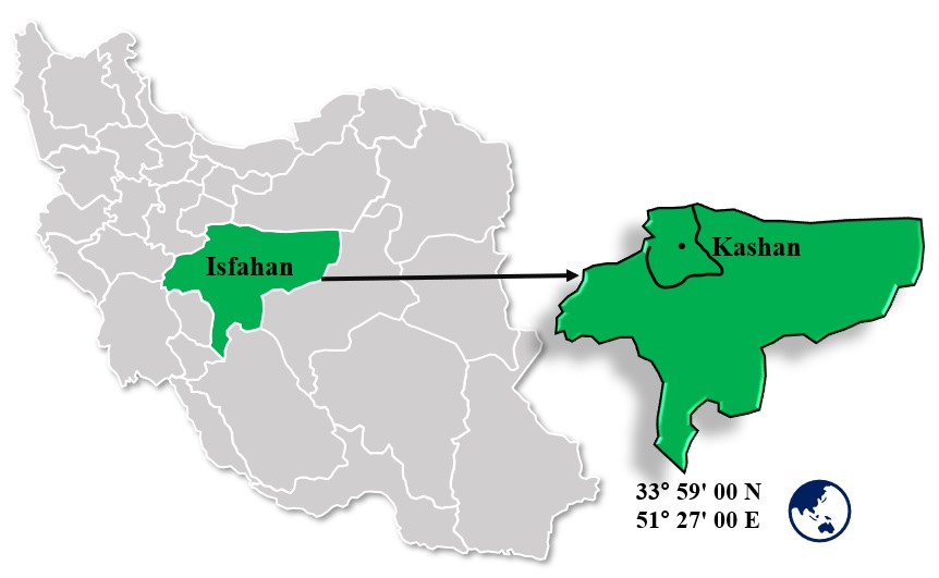
Fig.1. Map of Iran and Kashan
Table 1. Frequency Distribution of Demographic Variables and Risk Factors Associated with Hydatid Cyst in Study Participants
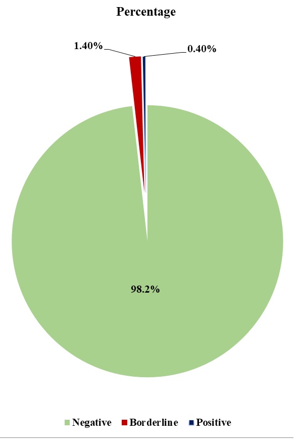
Fig. 2. Hydatid Cyst Seropositivity Rates in Rural Kashan: Overall Distribution
Table 2. Serological Test Results for Hydatid Cyst by Health Center in Rural Areas of Kashan
Table 2 outlines the overall seroprevalence of hydatid cyst among the 280 rural residents of Kashan County included in this study. A major share of participants tested negative for hydatid cyst antibodies, with 275 individuals (98.2%) receiving a negative result. A small proportion of the participants, 4 individuals (1.4%), showed borderline serological results. Only one participant (0.4%) tested positive for hydatid cyst antibodies.
The largest proportion of the study population originated from the Qamsar region (n=62, 22.1%), whereas the Barzok region accounted for the smallest number of participants (n=49, 17.5%). The only positive case was identified within the Qamsar region. Borderline cases were reported in the Joshaghan estark (n=1), Qamsar (n=1), and Joshaghan ghali (n=2) regions. In contrast, no borderline or positive cases were observed in other regions, including Barzok and Mahmoudabad.
Discussion
The findings of this study demonstrated a very low seroprevalence of hydatid cyst in the rural areas of Kashan County. There is a noticeable disparity between this finding with several national investigations, such as the one performed by Esmaeili et al. (2008) in Kashan [33], which indicated a prevalence of 3.05%. Further, when compared to other studies such as the research by Heydari et al. (2019) in Ardabil, which revealed a prevalence of 4.4% [32], the prevalence observed in the current study was lower. The variation in disease prevalence may emanate from improved sanitary conditions, enhanced public awareness, or geographical and lifestyle differences. Over time, increased public health awareness, ameliorated health literacy, and better sanitary practices have likely contributed to lowering exposure to the parasite [35]. In addition, changes in lifestyle, including diminished direct contact with dogs—the definitive host—and improved hygiene, may have further mitigated transmission.
Geographical variations in climate, environment, and socio-economic factors also play substantial roles in the differing prevalence rates [36]. Hydatid cyst prevalence varies widely in endemic regions such as the Mediterranean, Middle East, and parts of South America, suggesting the influence of regional health policies, cultural practices, and environmental conditions [37, 38].
Considering risk factors, this study found no statistically significant correlation between contact with dogs and serological test results (p = 0.142). This is in line with some previous studies that have identified contact with dogs as a potential risk factor for hydatid cyst infection [21, 39, 40]. In contrast, other variables, including gender, occupation, education level, vegetable washing method, and raw vegetable consumption, did not present a significant association with disease prevalence. These findings diverge from some similar research, such as the study by Darabi et al. (2020) in Arak, which reported a significant link between the type of vegetable washing and the prevalence rate [34]. This discrepancy may stem from variations in data collection methodologies or environmental factors.
The low prevalence noted in this study could be attributed to the improved level of public health and heightened public awareness regarding hydatid cyst prevention methods. In recent years, enhanced sanitary measures such as vegetable disinfection [41], animal vaccination, and improved control of livestock as well as stray dogs have likely had a significant influence on reducing the prevalence of this disease [39-41]. Also, the increased usage of detergents and improved vegetable washing practices may lower the risk of parasite transmission. Nevertheless, the results suggest that contact with infected dogs may still represent a risk factor that warrants further attention.
In studies carried out in other regions, the reported prevalence of the disease has been higher than the value observed in this investigation. For example, research in some rural areas of Iran has documented prevalence rates as high as 7.2 % [28]. This discrepancy may be attributed to environmental factors, variations in dietary habits, lifestyle differences, and even the genetic makeup of the studied populations. Moreover, the level of infection in livestock and dogs within the region can also affect the prevalence of the disease in humans.
Another significant factor that could contribute to discrepancies in reported prevalence rates across different studies is the type of diagnostic method utilized. Some studies have applied imaging techniques such as ultrasonography to directly identify hydatid cysts, while this study solely relied on a serological method (ELISA) for the detection of antibodies.
Although ELISA is a sensitive and specific method for detecting exposure to E. granulosus, it may not detect very early infections prior to antibody production, and false positives can occur owing to cross-reactivity with other parasitic infections. Meanwhile, serological testing enables identification of both symptomatic and asymptomatic cases that might be missed by imaging, especially in early or small cysts that are difficult to visualize.
Thus, although reliance on ELISA might underestimate some very recent infections, it provides a practical and reliable approach for large-scale screening in epidemiological surveys.
These findings suggest that the demographic and behavioral factors explored in this study are unlikely to have a direct and significant impact on the prevalence of hydatid cyst in the studied population. Nonetheless, it is plausible that other environmental or lifestyle-related factors not addressed here may contribute to disease transmission. Future studies with larger sample sizes, more diverse populations, and advanced statistical modeling are recommended to further clarify the complex determinants of hydatid cyst prevalence.
It is also suggested that future research focus on other potential factors, including individual hygiene and nutritional status, environmental impacts, and a more detailed examination of the association between contact with dogs and disease prevalence. Such studies could contribute to the development of more effective strategies for the control and prevention of this disease in rural communities.
Conclusion
This study indicated that the seroprevalence of hydatid cyst in rural areas of Kashan is relatively low. Even though contact with dogs was considered a potential risk factor, further studies with a larger sample size and more rigorous study designs are needed to confirm this association. Since hydatid cyst disease remains a significant public health and economic concern in some regions of Iran, it is recommended that educational and preventive programs be prioritized, including increasing public awareness about proper vegetable washing methods and reducing contact with infected dogs.
Acknowledgements
We would like to express our sincere gratitude to the distinguished Vice-Chancellor for Student Research Committee, Kashan University of Medical Sciences. Their invaluable and unwavering support was instrumental in the progress and completion of this research.
Conflict of interest
None declared.
Funding
This study was supported by Kashan University of Medical Sciences under grant number 402155.
Ethical Considerations
This study was conducted in accordance with the ethical principles of the Declaration of Helsinki for research involving human participants. Prior to the initiation of the study, the research protocol was reviewed and approved by the Ethics Committee of Kashan University of Medical Sciences. All participants were informed about the objectives, procedures, and the voluntary nature of the study, and written informed consent was obtained from each participant before sample collection. Participants were assured that their personal information would remain confidential and that all collected data would be anonymized and used solely for research purposes. Participants had the right to withdraw from the study at any stage without consequences. Blood sampling and data collection were performed by trained healthcare personnel in accordance with standard safety and hygiene protocols to minimize any potential risk to participants.
Code of Ethics
The study protocol was approved by the Ethics Committee of Kashan University of Medical Sciences (Ethics code: IR.KAUMS.MEDNT.REC.1402.279).
Authors' Contributions
Abolfazl Masoumi Koushk Mehdi and Mohammad Reza Shiee: designed and supervised the study. Data analysis and interpretation were carried out; Sina Karami and Farhat Sadat Firuzeh: were responsible for sample collection and field investigations in rural areas of Kashan; Mehdi Nazeri and Maryam Gharibi: performed the laboratory tests and contributed to data acquisition. All authors contributed to drafting and revising the manuscript and approved the final version for publication.
Full-Text: (14 Views)
Introduction
Hydatidosis, commonly referred to as hydatid cyst disease, is a zoonotic disease arising from human infection with the larval stage of Echinococcus granulosus [1]. This parasite is recognized as one of the most significant helminth infections transmitted between humans and animals [2]. The definitive host for this parasite is the dog, while livestock act as intermediate hosts. Humans inadvertently become infected as aberrant intermediate hosts [3]. The transmission of the parasite occurs between canids (definitive hosts that harbor the adult tapeworm stage in their intestines) and livestock, especially sheep (intermediate hosts that become infected through the fecal-oral route by ingesting eggs shed in dog feces). The mode of disease transmission to humans involves the ingestion of eggs excreted in dog feces via contaminated water or vegetables [4, 5]. The disease of cystic echinococcosis in humans is serious, chronic, and often overlooked [6]. The range of clinical manifestations varies from asymptomatic to severe and even life-threatening conditions [7]. It is common for asymptomatic or dormant cases to remain undiagnosed for years or even decades. Diagnosis of hydatid cysts in humans primarily relies on imaging techniques. Hydatid cysts in the abdomen are diagnosed via ultrasound imaging [8-10]. Diagnostic accuracy may be limited due to the use of crude antigens, stage‑dependent variability in sensitivity, the lack of standardized stage‑specific diagnostic methods, and the limited availability of rapid point‑of‑care tests.[11]. Serological tests for the detection of circulating antibodies in the serum of patients with hydatid cysts serve as a complementary tool [12, 13]. Currently, there is no commercially available circulating antigen detection tests [14, 15]. Hydatid cysts can develop in different organs of the body, though the liver is primarily affected, followed by the lungs as the most commonly involved organs. While virtually all organs and tissues can potentially host metacestodes, the most frequently affected organ is the liver (70%), followed by the lungs (20%), with less common involvement including the kidneys, spleen, muscles, and skin [16-18]. There are several treatment options for hydatid cysts, including surgical (conservative or radical), puncture-aspiration-injection-reaspiration (PAIR), antihelminthic drugs, and active surveillance [19, 20].
The infection caused by this parasite is endemic in many countries worldwide, including the West Asia region, the Mediterranean, South America, Iceland, Southern Africa, Australia, and New Zealand. The incidence of cysticcystic echinococcosis (CE) varies widely across different endemic regions, ranging from 1 to 220 cases per 1000000 population. These endemic areas include the Mediterranean countries, the Middle East, southern parts of South America, Iceland, Australia, New Zealand, and southern parts of Africa [21]. An estimated 6% pooled seroprevalence has been observed in different regions of Iran, ranging between 1.2% and 21%. In livestock and dogs, the prevalence is reported to be 6.73% and 5% to 49%, respectively. Further, the incidence of cases requiring surgical intervention is estimated at 1.2 per 100,000 population [22-25]. Hydatid cyst is considered a significant public health and economic issue across several developing countries, including Iran, where it is associated with high mortality. Moreover, it causes considerable financial burden on affected individuals. This disease affects approximately 1.2 million people worldwide, and its global burden is estimated at 188,000 new cases annually [26, 27]. Seroprevalence surveys have revealed asymptomatic cystic echinococcosis in 7.3% of individuals in rural areas of southeastern Kerman Province and 5.6% in voluntary blood donors from Fars Province in southern Iran [24, 28]. Based on the Iranian Health Ministry, the seroprevalence rates for hydatid cysts in some regions of Iran have been reported to be 3.4% in Alborz Province, 2.6% in Lorestan Province, 1.79 % in Meshginshahr, and 7.2% in Yasuj [29-32]. Even though several studies have been undertaken in other provinces, there is limited recent data on the seroprevalence of hydatid cysts in the rural population of Kashan. The last known seroepidemiological study in this region was performed by Esmaeili et al. in 2008 [33]. Since more than a decade has passed and given the likelihood of changes in environmental and behavioral risk factors, an updated study is essential. Our research aims to fill this gap through providing current, region-specific data that could inform public health planning and control measures in this endemic area.
The present study was undertaken to examine the seroprevalence of hydatid cyst and its associated risk factors among rural residents of Kashan County.
Materials and Methods
Kashan City is located in Center of Iran in Isfahan Province (fig.1), and surrounded by mountains and desert in the north, east, and south with a latitude of 33° 59' 00 N and longitude of 51° 27' 00 E. It is one of the main cities of the province whose population is 364,482 according to the last census in 2016, with at least one third of them living in the urban areas.
This study is a descriptive seroepidemiological cross-sectional survey carried out to determine the prevalence and associated risk factors of hydatid cyst among residents of rural areas in Kashan.
The sample size was calculated based on a previous study undertaken in Kashan by Esmaeili et al. (2008), which reported a seroprevalence of 3.05%. Assuming a 95% confidence level and a margin of error of 0.02, the required sample size was calculated by the following formula:
Formula 1.
n = (Z² × P × (1 – P)) / d²
Substituting the values:
Formula 2.
n = (1.962 × 0.0305 × 0.9695) / (0.02)2 ≈ 280
Thus, a total of 280 participants were included in the study. Initially, informed consent was obtained from these individuals, who were then questioned using a questionnaire regarding information relevant to this research. Next, 2 ml of blood was collected from each individual who agreed to participate in the study. Upon receipt of the sera from the laboratory, they were stored at -20°C in order ensure sample stability until the ELISA test was performed. These samples were gathered from five rural centers in Kashan, including Barzok, Joshaghan estark, Joshaghan ghali, Ghamsar, and Mohammadabad. Inclusion criteria were individuals with a fully functioning immune system residing in these areas. Exclusion criteria included individuals diagnosed with other parasitic diseases or those with immunodeficiency.
A questionnaire was completed for each individual for collecting information on various factors influencing the disease, such as age, sex, occupation, place of residence, literacy, etc. Children's parents, as well as participants, gave informed consent. This study was approved by the Ethics Committee of Kashan University of Medical
Sciences.
In order to determine the antibody status against hydatid cyst in individuals' serum samples, the Pishtaz Teb IgG anti-Echinococcus granulosus ELISA kit was employed. [34]. Following the kit's protocol, the cut-off optical density (OD) was specified using the negative control sample provided in the kit. Positive samples were defined as having an OD 10% above the cut-off, whereas negative samples were defined as having an OD 10% below the cut-off.
SPSS software version 16 was utilized for the entire data analysis; a P-value of less than 0.05 was deemed statistically significant.
Hydatidosis, commonly referred to as hydatid cyst disease, is a zoonotic disease arising from human infection with the larval stage of Echinococcus granulosus [1]. This parasite is recognized as one of the most significant helminth infections transmitted between humans and animals [2]. The definitive host for this parasite is the dog, while livestock act as intermediate hosts. Humans inadvertently become infected as aberrant intermediate hosts [3]. The transmission of the parasite occurs between canids (definitive hosts that harbor the adult tapeworm stage in their intestines) and livestock, especially sheep (intermediate hosts that become infected through the fecal-oral route by ingesting eggs shed in dog feces). The mode of disease transmission to humans involves the ingestion of eggs excreted in dog feces via contaminated water or vegetables [4, 5]. The disease of cystic echinococcosis in humans is serious, chronic, and often overlooked [6]. The range of clinical manifestations varies from asymptomatic to severe and even life-threatening conditions [7]. It is common for asymptomatic or dormant cases to remain undiagnosed for years or even decades. Diagnosis of hydatid cysts in humans primarily relies on imaging techniques. Hydatid cysts in the abdomen are diagnosed via ultrasound imaging [8-10]. Diagnostic accuracy may be limited due to the use of crude antigens, stage‑dependent variability in sensitivity, the lack of standardized stage‑specific diagnostic methods, and the limited availability of rapid point‑of‑care tests.[11]. Serological tests for the detection of circulating antibodies in the serum of patients with hydatid cysts serve as a complementary tool [12, 13]. Currently, there is no commercially available circulating antigen detection tests [14, 15]. Hydatid cysts can develop in different organs of the body, though the liver is primarily affected, followed by the lungs as the most commonly involved organs. While virtually all organs and tissues can potentially host metacestodes, the most frequently affected organ is the liver (70%), followed by the lungs (20%), with less common involvement including the kidneys, spleen, muscles, and skin [16-18]. There are several treatment options for hydatid cysts, including surgical (conservative or radical), puncture-aspiration-injection-reaspiration (PAIR), antihelminthic drugs, and active surveillance [19, 20].
The infection caused by this parasite is endemic in many countries worldwide, including the West Asia region, the Mediterranean, South America, Iceland, Southern Africa, Australia, and New Zealand. The incidence of cysticcystic echinococcosis (CE) varies widely across different endemic regions, ranging from 1 to 220 cases per 1000000 population. These endemic areas include the Mediterranean countries, the Middle East, southern parts of South America, Iceland, Australia, New Zealand, and southern parts of Africa [21]. An estimated 6% pooled seroprevalence has been observed in different regions of Iran, ranging between 1.2% and 21%. In livestock and dogs, the prevalence is reported to be 6.73% and 5% to 49%, respectively. Further, the incidence of cases requiring surgical intervention is estimated at 1.2 per 100,000 population [22-25]. Hydatid cyst is considered a significant public health and economic issue across several developing countries, including Iran, where it is associated with high mortality. Moreover, it causes considerable financial burden on affected individuals. This disease affects approximately 1.2 million people worldwide, and its global burden is estimated at 188,000 new cases annually [26, 27]. Seroprevalence surveys have revealed asymptomatic cystic echinococcosis in 7.3% of individuals in rural areas of southeastern Kerman Province and 5.6% in voluntary blood donors from Fars Province in southern Iran [24, 28]. Based on the Iranian Health Ministry, the seroprevalence rates for hydatid cysts in some regions of Iran have been reported to be 3.4% in Alborz Province, 2.6% in Lorestan Province, 1.79 % in Meshginshahr, and 7.2% in Yasuj [29-32]. Even though several studies have been undertaken in other provinces, there is limited recent data on the seroprevalence of hydatid cysts in the rural population of Kashan. The last known seroepidemiological study in this region was performed by Esmaeili et al. in 2008 [33]. Since more than a decade has passed and given the likelihood of changes in environmental and behavioral risk factors, an updated study is essential. Our research aims to fill this gap through providing current, region-specific data that could inform public health planning and control measures in this endemic area.
The present study was undertaken to examine the seroprevalence of hydatid cyst and its associated risk factors among rural residents of Kashan County.
Materials and Methods
Kashan City is located in Center of Iran in Isfahan Province (fig.1), and surrounded by mountains and desert in the north, east, and south with a latitude of 33° 59' 00 N and longitude of 51° 27' 00 E. It is one of the main cities of the province whose population is 364,482 according to the last census in 2016, with at least one third of them living in the urban areas.
This study is a descriptive seroepidemiological cross-sectional survey carried out to determine the prevalence and associated risk factors of hydatid cyst among residents of rural areas in Kashan.
The sample size was calculated based on a previous study undertaken in Kashan by Esmaeili et al. (2008), which reported a seroprevalence of 3.05%. Assuming a 95% confidence level and a margin of error of 0.02, the required sample size was calculated by the following formula:
Formula 1.
n = (Z² × P × (1 – P)) / d²
Substituting the values:
Formula 2.
n = (1.962 × 0.0305 × 0.9695) / (0.02)2 ≈ 280
Thus, a total of 280 participants were included in the study. Initially, informed consent was obtained from these individuals, who were then questioned using a questionnaire regarding information relevant to this research. Next, 2 ml of blood was collected from each individual who agreed to participate in the study. Upon receipt of the sera from the laboratory, they were stored at -20°C in order ensure sample stability until the ELISA test was performed. These samples were gathered from five rural centers in Kashan, including Barzok, Joshaghan estark, Joshaghan ghali, Ghamsar, and Mohammadabad. Inclusion criteria were individuals with a fully functioning immune system residing in these areas. Exclusion criteria included individuals diagnosed with other parasitic diseases or those with immunodeficiency.
A questionnaire was completed for each individual for collecting information on various factors influencing the disease, such as age, sex, occupation, place of residence, literacy, etc. Children's parents, as well as participants, gave informed consent. This study was approved by the Ethics Committee of Kashan University of Medical
Sciences.
In order to determine the antibody status against hydatid cyst in individuals' serum samples, the Pishtaz Teb IgG anti-Echinococcus granulosus ELISA kit was employed. [34]. Following the kit's protocol, the cut-off optical density (OD) was specified using the negative control sample provided in the kit. Positive samples were defined as having an OD 10% above the cut-off, whereas negative samples were defined as having an OD 10% below the cut-off.
SPSS software version 16 was utilized for the entire data analysis; a P-value of less than 0.05 was deemed statistically significant.
Fig.1. Map of Iran and Kashan
Results
In this study, 280 residents of rural areas in Kashan County were surveyed. The pie chart (Fig. 1) indicates that 98.2% of participants tested negative, 1.4% had borderline results, and only 0.4% tested positive for hydatid cyst antibodies based on serological testing. The following table reports the descriptive statistics of the demographic variables and other characteristics of the participants.
In this study, 280 residents of rural areas in Kashan County were surveyed. The pie chart (Fig. 1) indicates that 98.2% of participants tested negative, 1.4% had borderline results, and only 0.4% tested positive for hydatid cyst antibodies based on serological testing. The following table reports the descriptive statistics of the demographic variables and other characteristics of the participants.
Table 1. Frequency Distribution of Demographic Variables and Risk Factors Associated with Hydatid Cyst in Study Participants
| Variable |
| Frequency (Percentage) | ||
| Gender | Male | 89 (31.8%) |
| Female | 191 (68.2%) | |
| Age groups | Under 35 years | 67 (23.9%) |
| 35–50 years | 69 (24.6%) | |
| 51–65 years | 75 (26.8%) | |
| Over 65 years | 69 (24.6%) | |
| Education level | Illiterate | 82 (29.9%) |
| Primary school | 87 (31.8%) | |
| Middle school | 56 (20.4%) | |
| High school | 30 (10.9%) | |
| University degree | 19 (6.9%) | |
| Occupation | Farmer, rancher, laborer | 14 (5.8%) |
| Housewife | 146 (60.3%) | |
| Employee | 22 (9.1%) | |
| Self-employed | 40 (16.5%) | |
| Retired | 20 (8.3%) | |
| Vegetable washing method | Salt water | 158 (57.2%) |
| Detergent | 51 (18.5%) | |
| Water/Vinegar/Baking soda | 67 (24.3%) | |
| Contact with dogs | Yes | 43 (15.4%) |
| No | 236 (84.6%) | |
| Serological results | Negative | 275 (98.2%) |
| Doubtful | 4 (1.4%) | |
| Positive | 1 (0.4%) |
The descriptive statistics (Table 1) demonstrated that the majority of participants in this study were female (68.2%). The mean age of the individuals was 49.6 years, with the 51–65-year age group representing the largest proportion. Considering education level, the highest frequency was observed at the primary level (31.8%), whereas the lowest was at the university level (6.9%).
With regards to the occupational status, it was found that housewives constituted the largest group of participants (60.3%), while farmers and livestock
breeders represented the smallest group (5.8%). In terms of lifestyle factors, 86% of individuals reported consuming raw vegetables, and 15.4% had a history of contact with dogs. Considering vegetable washing methods, the majority of individuals (57.2%) utilized water and salt. Based on the serological test results, 98.2% of individuals had a negative result, while 1.4% were suspect and 0.4% were positive (Fig. 2). These data reflect a low prevalence of hydatid cyst in this region; however, factors such as contact with dogs and inadequate food washing practices may be involved in the spread of the disease.
With regards to the occupational status, it was found that housewives constituted the largest group of participants (60.3%), while farmers and livestock
breeders represented the smallest group (5.8%). In terms of lifestyle factors, 86% of individuals reported consuming raw vegetables, and 15.4% had a history of contact with dogs. Considering vegetable washing methods, the majority of individuals (57.2%) utilized water and salt. Based on the serological test results, 98.2% of individuals had a negative result, while 1.4% were suspect and 0.4% were positive (Fig. 2). These data reflect a low prevalence of hydatid cyst in this region; however, factors such as contact with dogs and inadequate food washing practices may be involved in the spread of the disease.
Fig. 2. Hydatid Cyst Seropositivity Rates in Rural Kashan: Overall Distribution
The chi-square test results reveal that none of the examined variables presented a statistically significant association with the serological test outcomes, as the p-value for all variables was greater than 0.05. This implies that gender, occupation, education level, vegetable washing method, and raw vegetable consumption did not have a significant influence on the likelihood of hydatid cyst infection. The only variable, contact with dogs, with a p-value of 0.142, was close to the significance threshold. This suggests that this variable may have a potential effect on the disease prevalence with a larger sample size or the usage of more advanced statistical models.
Table 2. Serological Test Results for Hydatid Cyst by Health Center in Rural Areas of Kashan
| Health Center | Negative (n/%) | Borderline (n/%) | Positive (n/%) | Total (n/%) |
| Barzok | 49 (17.5%) | 0 (0%) | 0 (0%) | 49 (17.5%) |
| Joshaghan estark | 55 (19.6%) | 1 (0.4%) | 0 (0%) | 56 (20%) |
| Mahmoodabad | 61 (21.8%) | 0 (0%) | 0 (0%) | 61 (21.8%) |
| Ghamsar | 60 (21.4%) | 1 (0.4%) | 1 (0.4%) | 62 (22.1%) |
| Joshaghan ghali | 50 (17.9%) | 2 (0.7%) | 0 (0%) | 52 (18.6%) |
| Total | 275 (98.2%) | 4 (1.4%) | 1 (0.4%) |
| 280 (100%) |
Table 2 outlines the overall seroprevalence of hydatid cyst among the 280 rural residents of Kashan County included in this study. A major share of participants tested negative for hydatid cyst antibodies, with 275 individuals (98.2%) receiving a negative result. A small proportion of the participants, 4 individuals (1.4%), showed borderline serological results. Only one participant (0.4%) tested positive for hydatid cyst antibodies.
The largest proportion of the study population originated from the Qamsar region (n=62, 22.1%), whereas the Barzok region accounted for the smallest number of participants (n=49, 17.5%). The only positive case was identified within the Qamsar region. Borderline cases were reported in the Joshaghan estark (n=1), Qamsar (n=1), and Joshaghan ghali (n=2) regions. In contrast, no borderline or positive cases were observed in other regions, including Barzok and Mahmoudabad.
Discussion
The findings of this study demonstrated a very low seroprevalence of hydatid cyst in the rural areas of Kashan County. There is a noticeable disparity between this finding with several national investigations, such as the one performed by Esmaeili et al. (2008) in Kashan [33], which indicated a prevalence of 3.05%. Further, when compared to other studies such as the research by Heydari et al. (2019) in Ardabil, which revealed a prevalence of 4.4% [32], the prevalence observed in the current study was lower. The variation in disease prevalence may emanate from improved sanitary conditions, enhanced public awareness, or geographical and lifestyle differences. Over time, increased public health awareness, ameliorated health literacy, and better sanitary practices have likely contributed to lowering exposure to the parasite [35]. In addition, changes in lifestyle, including diminished direct contact with dogs—the definitive host—and improved hygiene, may have further mitigated transmission.
Geographical variations in climate, environment, and socio-economic factors also play substantial roles in the differing prevalence rates [36]. Hydatid cyst prevalence varies widely in endemic regions such as the Mediterranean, Middle East, and parts of South America, suggesting the influence of regional health policies, cultural practices, and environmental conditions [37, 38].
Considering risk factors, this study found no statistically significant correlation between contact with dogs and serological test results (p = 0.142). This is in line with some previous studies that have identified contact with dogs as a potential risk factor for hydatid cyst infection [21, 39, 40]. In contrast, other variables, including gender, occupation, education level, vegetable washing method, and raw vegetable consumption, did not present a significant association with disease prevalence. These findings diverge from some similar research, such as the study by Darabi et al. (2020) in Arak, which reported a significant link between the type of vegetable washing and the prevalence rate [34]. This discrepancy may stem from variations in data collection methodologies or environmental factors.
The low prevalence noted in this study could be attributed to the improved level of public health and heightened public awareness regarding hydatid cyst prevention methods. In recent years, enhanced sanitary measures such as vegetable disinfection [41], animal vaccination, and improved control of livestock as well as stray dogs have likely had a significant influence on reducing the prevalence of this disease [39-41]. Also, the increased usage of detergents and improved vegetable washing practices may lower the risk of parasite transmission. Nevertheless, the results suggest that contact with infected dogs may still represent a risk factor that warrants further attention.
In studies carried out in other regions, the reported prevalence of the disease has been higher than the value observed in this investigation. For example, research in some rural areas of Iran has documented prevalence rates as high as 7.2 % [28]. This discrepancy may be attributed to environmental factors, variations in dietary habits, lifestyle differences, and even the genetic makeup of the studied populations. Moreover, the level of infection in livestock and dogs within the region can also affect the prevalence of the disease in humans.
Another significant factor that could contribute to discrepancies in reported prevalence rates across different studies is the type of diagnostic method utilized. Some studies have applied imaging techniques such as ultrasonography to directly identify hydatid cysts, while this study solely relied on a serological method (ELISA) for the detection of antibodies.
Although ELISA is a sensitive and specific method for detecting exposure to E. granulosus, it may not detect very early infections prior to antibody production, and false positives can occur owing to cross-reactivity with other parasitic infections. Meanwhile, serological testing enables identification of both symptomatic and asymptomatic cases that might be missed by imaging, especially in early or small cysts that are difficult to visualize.
Thus, although reliance on ELISA might underestimate some very recent infections, it provides a practical and reliable approach for large-scale screening in epidemiological surveys.
These findings suggest that the demographic and behavioral factors explored in this study are unlikely to have a direct and significant impact on the prevalence of hydatid cyst in the studied population. Nonetheless, it is plausible that other environmental or lifestyle-related factors not addressed here may contribute to disease transmission. Future studies with larger sample sizes, more diverse populations, and advanced statistical modeling are recommended to further clarify the complex determinants of hydatid cyst prevalence.
It is also suggested that future research focus on other potential factors, including individual hygiene and nutritional status, environmental impacts, and a more detailed examination of the association between contact with dogs and disease prevalence. Such studies could contribute to the development of more effective strategies for the control and prevention of this disease in rural communities.
Conclusion
This study indicated that the seroprevalence of hydatid cyst in rural areas of Kashan is relatively low. Even though contact with dogs was considered a potential risk factor, further studies with a larger sample size and more rigorous study designs are needed to confirm this association. Since hydatid cyst disease remains a significant public health and economic concern in some regions of Iran, it is recommended that educational and preventive programs be prioritized, including increasing public awareness about proper vegetable washing methods and reducing contact with infected dogs.
Acknowledgements
We would like to express our sincere gratitude to the distinguished Vice-Chancellor for Student Research Committee, Kashan University of Medical Sciences. Their invaluable and unwavering support was instrumental in the progress and completion of this research.
Conflict of interest
None declared.
Funding
This study was supported by Kashan University of Medical Sciences under grant number 402155.
Ethical Considerations
This study was conducted in accordance with the ethical principles of the Declaration of Helsinki for research involving human participants. Prior to the initiation of the study, the research protocol was reviewed and approved by the Ethics Committee of Kashan University of Medical Sciences. All participants were informed about the objectives, procedures, and the voluntary nature of the study, and written informed consent was obtained from each participant before sample collection. Participants were assured that their personal information would remain confidential and that all collected data would be anonymized and used solely for research purposes. Participants had the right to withdraw from the study at any stage without consequences. Blood sampling and data collection were performed by trained healthcare personnel in accordance with standard safety and hygiene protocols to minimize any potential risk to participants.
Code of Ethics
The study protocol was approved by the Ethics Committee of Kashan University of Medical Sciences (Ethics code: IR.KAUMS.MEDNT.REC.1402.279).
Authors' Contributions
Abolfazl Masoumi Koushk Mehdi and Mohammad Reza Shiee: designed and supervised the study. Data analysis and interpretation were carried out; Sina Karami and Farhat Sadat Firuzeh: were responsible for sample collection and field investigations in rural areas of Kashan; Mehdi Nazeri and Maryam Gharibi: performed the laboratory tests and contributed to data acquisition. All authors contributed to drafting and revising the manuscript and approved the final version for publication.
References
1. Mohammed MM, Oreibi RA. Hydatidosis and its effects on humans and animals. [M.Sc. thesis]. Republic of Iraq: University of Al-Qadisiyah; 2021.
2. Wells K, Gibson DI, Clark NJ, Ribas A, Morand S, McCallum HI. Global spread of helminth parasites at the human–domestic animal–wildlife interface. Glob Chang Biol. 2018;24(7):3254-65. [DOI] [PMID]
3. Agudelo Higuita NI, Brunetti E, McCloskey C. Cystic Echinococcosis. J Clin Microbiol. 2016;54(3):518-23. [DOI] [PMID] [PMCID]
4. Craig PS, McManus DP, Lightowlers MW, Chabalgoity JA, Garcia HH, Gavidia CM, et al. Prevention and control of cystic echinococcosis. Lancet Infect Dis. 2007;7(6):385-94. [DOI] [PMID]
5. da Silva AM. Human echinococcosis: a neglected disease. Gastroenterol Res Pract. 2010;2010:583297. [DOI] [PMID] [PMCID]
6. Brunetti E, Garcia HH, Junghanss T; International CE Workshop in Lima, Peru, 2009. Cystic echinococcosis: chronic, complex, and still neglected. PLoS Negl Trop Dis. 2011;5(7):e1146. [DOI] [PMID] [PMCID]
7. Khalili N, Iranpour P, Khalili N, Haseli S. Hydatid Disease: A Pictorial Review of Uncommon Locations. Iran J Med Sci. 2023;48(2):118-29. [DOI] [PMID] [PMCID]
8. Joshi U, Subedi R, Jayswal A, Agrawal V. Clinical Characteristics and Management of the Hydatid Cyst of the Liver: A Study from a Tertiary Care Center in Nepal. J Parasitol Res. 2020;2020:8867744. [DOI] [PMID] [PMCID]
9. Stojkovic M, Rosenberger K, Kauczor HU, Junghanss T, Hosch W. Diagnosing and staging of cystic echinococcosis: how do CT and MRI perform in comparison to ultrasound? PLoS Negl Trop Dis. 2012;6(10):e1880. [DOI] [PMID] [PMCID]
10. 10. Mohemmi N, Khodayari MT, Karamati SA, Shiee MR, Effatpanah H, Davari A, et al. Update on the Seroepidemiology of Human Cystic Echinococcosis and Associated Risk Factors in Iran: A Systematic Review and Meta-Analysis. Health Sci Rep. 2025;8(4):e70699. [DOI] [PMID] [PMCID]
11. Siles-Lucas M, Casulli A, Conraths FJ, Müller N. Laboratory Diagnosis of Echinococcus spp. in Human Patients and Infected Animals. Adv Parasitol. 2017;96:159-257. [DOI] [PMID]
12. Vola A, Manciulli T, De Silvestri A, Lissandrin R, Mariconti M, Siles-Lucas M, et al. Diagnostic Performances of Commercial ELISA, Indirect Hemagglutination, and Western Blot in Differentiation of Hepatic Echinococcal and Non-Echinococcal Lesions: A Retrospective Analysis of Data from a Single Referral Centre. Am J Trop Med Hyg. 2019;101(6):1345-9. [DOI] [PMID] [PMCID]
13. Eckert J, Deplazes P. Biological, epidemiological, and clinical aspects of echinococcosis, a zoonosis of increasing concern. Clin Microbiol Rev. 2004;17(1):107-35. [DOI] [PMID] [PMCID]
14. Bresson-Hadni S, Spahr L, Chappuis F. Hepatic Alveolar Echinococcosis. Semin Liver Dis. 2021;41(3):393-408. [DOI] [PMID]
15. Tsaroucha AK, Polychronidis AC, Lyrantzopoulos N, Pitiakoudis MS, J Karayiannakis A,, Manolas KJ, et al. Hydatid disease of the abdomen and other locations. World J Surg. 2005;29(9):1161-5. [DOI] [PMID]
16. Mihmanli M, Idiz UO, Kaya C, Demir U, Bostanci O, Omeroglu S, et al. Current status of diagnosis and treatment of hepatic echinococcosis. World J Hepatol. 2016;8(28):1169-81. [DOI] [PMID] [PMCID]
17. Ramia Ángel JM, Manuel Vázquez A, Gijón Román C, Latorre Fragua R, de la Plaza Llamas R. Radical surgery in hepatic hydatidosis: analysis of results in an endemic area. Rev Esp Enferm Dig. 2020;112(9):708-11. [DOI] [PMID]
18. Gessese AT. Review on Epidemiology and Public Health Significance of Hydatidosis. Vet Med Int. 2020;2020:8859116. [DOI] [PMID] [PMCID]
19. Dalimi A, Motamedi G, Hosseini M, Mohammadian B, Malaki H, Ghamari Z, et al. Echinococcosis/hydatidosis in western Iran. Vet Parasitol. 2002;105(2):161-71. [DOI] [PMID]
20. Galeh TM, Spotin A, Mahami-Oskouei M, Carmena D, Rahimi MT, Barac A, et al. The seroprevalence rate and population genetic structure of human cystic echinococcosis in the Middle East: A systematic review and meta-analysis. Int J Surg. 2018;51:39-48. [DOI] [PMID]
21. Sarkari B, Hosseini F, Abdolahi Khabisi S, Sedaghat F. Seroprevalence of cystic echinococcosis in blood donors in Fars province, southern Iran. Parasite Epidemiol Control. 2017;2(1):8-12. [DOI] [PMID] [PMCID]
22. Shafiei R, Teshnizi SH, Kalantar K, Gholami M, Mirzaee G, Mirzaee F. The Seroprevalence of Human Cystic Echinococcosis in Iran: A Systematic Review and Meta-Analysis Study. J Parasitol Res. 2016;2016:1425147. [DOI] [PMID] [PMCID]
23. Kern P, Menezes da Silva A, Akhan O, Müllhaupt B, Vizcaychipi KA, Budke C,et al. The Echinococcoses: Diagnosis, Clinical Management and Burden of Disease. Adv Parasitol. 2017;96:259-369. [DOI] [PMID]
24. Deplazes P, Rinaldi L, Alvarez Rojas CA, Torgerson PR, Harandi MF, Romig T, et al. Global Distribution of Alveolar and Cystic Echinococcosis. Adv Parasitol. 2017;95:315-493. [DOI] [PMID]
25. Harandi MF, Moazezi SS, Saba M, Grimm F, Kamyabi H, Sheikhzadeh F, et al. Sonographical and serological survey of human cystic echinococcosis and analysis of risk factors associated with seroconversion in rural communities of Kerman, Iran. Zoonoses Public Health. 2011;58(8):582-8. [DOI] [PMID]
26. Sarkari B, Sadjjadi SM, Beheshtian MM, Aghaee M, Sedaghat F. Human cystic echinococcosis in Yasuj District in Southwest of Iran: an epidemiological study of seroprevalence and surgical cases over a ten-year period. Zoonoses Public Health. 2010;57(2):146-50. [DOI] [PMID]
27. Dabaghzadeh H, Bairami A, Kia EB, Aryaeipour M, Rokni MB. Seroprevalence of Human Cystic Echinococcosis in Alborz Province, Central Iran in 2015. Iran J Public Health. 2018;47(4):561-6. [PMID] [PMCID]
28. Fallah Omrani V, Rouhani S, Kazemi B, Seyyedtabaei SJ, Kheirandish F, Rezapour M. Seroprevalence of IgG Antibodies against Echinococcus granulosus by ELISA Method Using Recombinant Agb in Lorestan Province, Western Iran. Iran J Public Health. 2017;46(8):1132-8. [PMID] [PMCID]
29. Heidari Z, Mohebali M, Zarei Z, Aryayipour M, Eshraghian M, Kia E, et al. Seroepidemiological study of human hydatidosis in meshkinshahr district, ardabil province, Iran. Iran J Parasitol. 2011;6(3):19-25.
30. Esmaeili N, Arbabi M. Seroepidemiology of hydatidosis among adult human at Kashan region, Iran in 2008. Feyz Med Sci J. 2010;13(4):321-6. [URL]
31. Darabi F, Bakhtiari M, Matini S, Matini M. Seroepidemiology of Hydatid Cyst in Outpatients Attending Health Centers in Arak City, Iran, 2020. Avicenna J Clin Med. 2022. [DOI]
32. Hassan AH. Epidemiological Characteristics of Hydatid Cyst Disease Crucial for Targeted Public Health Interventions in Endemic Areas. Integr Biomed Res. 2024;8(6):1-6. [DOI]
33. Ghatee MA, Nikaein K, Taylor WR, Karamian M, Alidadi H, Kanannejad Z, et al. Environmental, climatic and host population risk factors of human cystic echinococcosis in southwest of Iran. BMC Public Health. 2020;20(1):1611. [DOI] [PMID] [PMCID]
34. Dakkak A. Echinococcosis/hydatidosis: a severe threat in Mediterranean countries. Vet Parasitol. 2010;174(1-2):2-11. [DOI] [PMID]
35. Grosso G, Gruttadauria S, Biondi A, Marventano S, Mistretta A. Worldwide epidemiology of liver hydatidosis including the Mediterranean area. World J Gastroenterol. 2012;18(13):1425-37. [DOI] [PMID] [PMCID]
36. Possenti A, Manzano-Román R, Sánchez-Ovejero C, Boufana B, La Torre G, Siles-Lucas M, et al. Potential Risk Factors Associated with Human Cystic Echinococcosis: Systematic Review and Meta-analysis. PLoS Negl Trop Dis. 2016;10(11):e0005114. [DOI] [PMID] [PMCID]
37. Acosta-Jamett G, Weitzel T, Boufana B, Adones C, Bahamonde A, Abarca K, et al. Prevalence and risk factors for echinococcal infection in a rural area of northern Chile: a household-based cross-sectional study. PLoS Negl Trop Dis. 2014;8(8):e3090. [DOI] [PMID] [PMCID]
38. Hajipour N, Soltani M, Ketzis J, Hassanzadeh P. Zoonotic parasitic organisms on vegetables: Impact of production system characteristics on presence, prevalence on vegetables in northwestern Iran and washing methods for removal. Food Microbiol. 2021;95:103704. [DOI] [PMID]
39. Khan A, Ahmed H, Simsek S, Afzal MS, Cao J. Spread of Cystic Echinococcosis in Pakistan Due to Stray Dogs and Livestock Slaughtering Habits: Research Priorities and Public Health Importance. Front Public Health. 2020;7:412. [DOI] [PMID] [PMCID]
Send email to the article author
| Rights and permissions | |
 |
This work is licensed under a Creative Commons Attribution 4.0 International License. |