Journal of Occupational
Health and Epidemiology

Rafsanjan university Of medical sciences
Volume 15, Issue 1 (Winter 2026)
J Occup Health Epidemiol 2026, 15(1): 78-88 |
Back to browse issues page



Ethics code: IR.AJUMS.REC.1404.117


Download citation:
BibTeX | RIS | EndNote | Medlars | ProCite | Reference Manager | RefWorks
Send citation to:


BibTeX | RIS | EndNote | Medlars | ProCite | Reference Manager | RefWorks
Send citation to:
Tabanfar S, Fouladi Dehaghi B, Sobhani S. The Impact of Radiofrequency Exposure on Reproductive Health in Mice and Rats: A Systematic Review. , J Occup Health Epidemiol. 2026; 15 (1) :78-88
URL: http://johe.rums.ac.ir/article-1-1062-en.html
URL: http://johe.rums.ac.ir/article-1-1062-en.html
Related article in
Google Scholar
Google Scholar
Similar articles


1- Ph.D. Student in Occupational Health and Safety, Dept. of Occupational Safety and Health Engineering, Faculty of Health, Student Research Committee, Ahvaz Jundishapur University of Medical Sciences, Ahvaz, Iran.
2- Professor, Dept. of Occupational Safety and Health Engineering, Faculty of Health, Ahvaz Jundishapur University of Medical Sciences, Ahvaz, Iran.
3- Ph.D. Student in Occupational Health and Safety, Dept. of Occupational Safety and Health Engineering, Faculty of Health, Student Research Committee, Ahvaz Jundishapur University of Medical Sciences, Ahvaz, Iran. ,seyvan.sobhani21@gmail.com
2- Professor, Dept. of Occupational Safety and Health Engineering, Faculty of Health, Ahvaz Jundishapur University of Medical Sciences, Ahvaz, Iran.
3- Ph.D. Student in Occupational Health and Safety, Dept. of Occupational Safety and Health Engineering, Faculty of Health, Student Research Committee, Ahvaz Jundishapur University of Medical Sciences, Ahvaz, Iran. ,
Article history

Received: 2025/06/20
Accepted: 2025/09/14
ePublished: 2026/03/30
Accepted: 2025/09/14
ePublished: 2026/03/30
Subject:
Occupational Health
Keywords: Radiofrequency [MeSH], Oxidative Stress [MeSH], Reproduction [MeSH], Non-Ionizing Radiation [MeSH]
Full-Text [PDF 477 kb]
(392 Downloads)
| Abstract (HTML) (1324 Views)
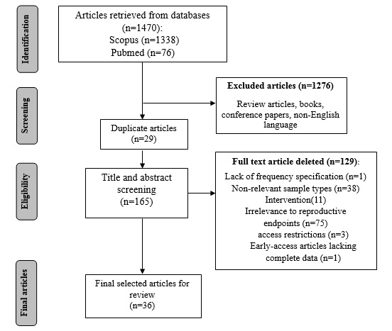
Fig. 1. The process of search, screening, and article selection according to the PRISMA protocol
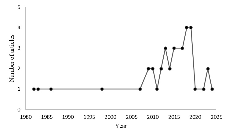
Fig. 2. Graph presenting the number of studies included in this review, categorized by publication year
Full-Text: (9 Views)
Introduction
The technological applications of radiofrequency electromagnetic fields (RF-EMF), encompassing the frequency spectrum from 100 kHz to 300 GHz, have indicated consistent growth since the mid-20th century [1]. RF-EMF is applicable across diverse domains including medical, industrial processes, domestic appliances (notably Wi-Fi systems), security and navigation technologies, with particular prominence in telecommunication sectors such as broadcasting and mobile telephony [2]. In the contemporary society, the imperative for continuous connectivity has culminated in ubiquitous exposure to non-ionizing radiation emitted by RF-EMF sources [3, 4]. Of paramount concern within this paradigm is the potential bioeffects of such electromagnetic exposure on human physiological systems.
In recent decades, growing concerns have emerged considering the global decline in human fertility rates. Among various environmental factors, exposure to radiofrequency (RF) fields has been proposed as a potential contributing risk factor [5]. Recognizing the significance of this issue, the World Health Organization (WHO) undertook a comprehensive international expert survey in 2018 to appraise research priorities regarding RF effects. The survey identified adverse reproductive outcomes as a top-tier research priority, with 32% consensus among respondents [1]. Although no significant clinical effects have been linked to RF exposure so far, numerous studies suggest that this type of energy may have subtle harmful effects on biological systems [6].
The biological impacts of RF-EMF emitted by wireless devices are broadly categorized into:
Thermal effects arising from localized heat generation during prolonged mobile phone use in close proximity to the body, and;
Non-thermal effects primarily mediated through generation of reactive oxygen species (ROS) and subsequent oxidative stress induction.
Both mechanisms have been implicated in germ cell cycle disruption [7]. Current evidence from epidemiological and experimental studies exploring RF-EMF effects on the reproductive system of laboratory animals, as well as fetal development, remains inconclusive [4]. While substantial research suggests that RF exposure may compromise male reproductive health through multiple pathways - including impaired sperm parameters (count, motility, morphology), histological alterations in seminiferous tubules, and subsequent fertility decline in both human and animal models [8] - similar concerns exist regarding female reproductive outcomes. Indeed, associations have been reported between RF exposure and increased risks of spontaneous abortion, congenital abnormalities, intrauterine growth restriction, and preterm delivery [9-11].
Notably, some studies have documented null effects of RF exposure on either male or female fertility parameters [3, 5, 12], highlighting the need for further standardized research in this field.
The growing use of RF-emitting devices such as mobile phones, Wi-Fi routers, base stations, and microwave ovens has created an urgent need to address their influence on human health. Maximum permissible exposure limits vary widely [13]. For example, the permissible Specific Absorption Rate (SAR) limits vary significantly across organizations. The Federal Communications Commission (FCC) sets a SAR limit of 1.6 W/kg, while the International Commission on Non-Ionizing Radiation Protection (ICNIRP) adopts a slightly higher limit of 2 W/kg. Both organizations base their standards primarily on short-term thermal effects. In contrast, countries such as Russia and China enforce limits up to ten times stricter, incorporating non-thermal effects into their standards. Notably, IARC classifies RF radiation as 'possibly carcinogenic' (Group 2B), a conclusion not reflected in current regulatory standards [14]. There is no definitive standard for the level and duration of RF exposure that induces adverse health effects, and this remains a challenging issue [14, 15]. Nevertheless, the existing literature on the effects of non-ionizing RF radiation reveals conflicting results of its biological and adverse health effects. The aim of this systematic review is to inspect current knowledge about the risks of radiofrequency waves on reproductive health in both males and females based on in vivo studies. We also summarize available evidence and appraise whether the literature supports or refutes the effects of RF exposure on reproductive health. These findings may be practical for developing new precautionary guidelines for exposure limits.
Materials and Methods
This systematic review explores the relationship between radiofrequency wave exposure and reproductive health in mice/ rats (male and female). We followed the Preferred Reporting Items for Systematic Reviews and Meta-Analyses (PRISMA) protocol for article search, analysis, evaluation, and summarization. The search was conducted in two stages.
In the first stage, we searched the ISI Web of Science, MEDLINE-PubMed, and Scopus databases using the following keywords: radiofrequency radiation, electromagnetic field, reproductive, fertility, infertility, testis, testicular, embryo, males, female, rats, mice, oxidative stress, sperm parameters, ROS, Wi-Fi, and phone. We utilized diverse keyword combinations, Boolean operators, and database-specific filters to conduct a comprehensive search. The search covered the entire available timeline in each database (from the inception of online indexing: 1900 for Web of Science, 1966 for PubMed, and 1970 for Scopus) up to August 2024, with no additional time restrictions applied. Searches employed Medical Subject Headings (MeSH). Following initial screening of titles and abstracts based on study objectives and criteria, we identified 165 eligible articles.
The second stage involved independent full-text review by the authors to select final articles. Through this process, 36 articles were identified for final inclusion. The full text of the identified studies was reviewed independently by two researchers. Disagreements were often resolved through discussion. If disagreements were not resolved through discussion, a third researcher was consulted. Fig. 1 displays the complete PRISMA-based search, screening, and selection process.
Inclusion and Exclusion Criteria: This systematic review included original articles published in English that had explored the effects of radiofrequency exposure on reproductive health in rats or mice. Eligible studies needed to be in vivo experiments reporting quantitative data on at least one reproductive endpoint (e.g., sperm parameters, hormonal levels, histopathological changes) with explicit documentation of RF exposure parameters (frequency, duration, SAR, Exposure dose). In vitro studies, computational models, human epidemiological studies, and research involving mammalian species other than mice and rats were excluded. Moreover, studies were excluded if they lacked control groups, represented duplicate publications, had no available full texts, or provided insufficient methodological details. Only peer-reviewed original articles were included in this review; other publication types (e.g., reviews, conference proceedings, books, and theses) were excluded.
Of the studies identified, eligibility for inclusion was determined through applying the Population, Exposure, Comparator, Outcomes (PECO) framework. The review
considered in vivo studies utilizing mouse or rat models (Population) that had inspected the effects of exposure to RF-EMF at any frequency or duration (Exposure). These studies were required to include a sham-exposed or completely unexposed control group for comparison (Comparator). The primary outcomes of interest covered a range of reproductive endpoints, including but not limited to, sperm quality parameters, serum hormonal levels, and histopathological alterations in reproductive tissues (Outcomes).
In addition to peer-reviewed studies, we also screened major sources of grey literature such as WHO/ICNIRP reports, national agency reports (e.g., SSM, ANSES), and the OpenGrey archive. Nevertheless, no additional primary animal studies relevant to this work were identified. Thus, our systematic review was based on peer-reviewed publications only.
Data Extraction: The following information was extracted from the included articles:
Study characteristics: Author, year, species, strain, gender
Exposure parameters: Frequency, specific absorption rate (SAR), duration, exposure setup (whole-body/localized)
The technological applications of radiofrequency electromagnetic fields (RF-EMF), encompassing the frequency spectrum from 100 kHz to 300 GHz, have indicated consistent growth since the mid-20th century [1]. RF-EMF is applicable across diverse domains including medical, industrial processes, domestic appliances (notably Wi-Fi systems), security and navigation technologies, with particular prominence in telecommunication sectors such as broadcasting and mobile telephony [2]. In the contemporary society, the imperative for continuous connectivity has culminated in ubiquitous exposure to non-ionizing radiation emitted by RF-EMF sources [3, 4]. Of paramount concern within this paradigm is the potential bioeffects of such electromagnetic exposure on human physiological systems.
In recent decades, growing concerns have emerged considering the global decline in human fertility rates. Among various environmental factors, exposure to radiofrequency (RF) fields has been proposed as a potential contributing risk factor [5]. Recognizing the significance of this issue, the World Health Organization (WHO) undertook a comprehensive international expert survey in 2018 to appraise research priorities regarding RF effects. The survey identified adverse reproductive outcomes as a top-tier research priority, with 32% consensus among respondents [1]. Although no significant clinical effects have been linked to RF exposure so far, numerous studies suggest that this type of energy may have subtle harmful effects on biological systems [6].
The biological impacts of RF-EMF emitted by wireless devices are broadly categorized into:
Thermal effects arising from localized heat generation during prolonged mobile phone use in close proximity to the body, and;
Non-thermal effects primarily mediated through generation of reactive oxygen species (ROS) and subsequent oxidative stress induction.
Both mechanisms have been implicated in germ cell cycle disruption [7]. Current evidence from epidemiological and experimental studies exploring RF-EMF effects on the reproductive system of laboratory animals, as well as fetal development, remains inconclusive [4]. While substantial research suggests that RF exposure may compromise male reproductive health through multiple pathways - including impaired sperm parameters (count, motility, morphology), histological alterations in seminiferous tubules, and subsequent fertility decline in both human and animal models [8] - similar concerns exist regarding female reproductive outcomes. Indeed, associations have been reported between RF exposure and increased risks of spontaneous abortion, congenital abnormalities, intrauterine growth restriction, and preterm delivery [9-11].
Notably, some studies have documented null effects of RF exposure on either male or female fertility parameters [3, 5, 12], highlighting the need for further standardized research in this field.
The growing use of RF-emitting devices such as mobile phones, Wi-Fi routers, base stations, and microwave ovens has created an urgent need to address their influence on human health. Maximum permissible exposure limits vary widely [13]. For example, the permissible Specific Absorption Rate (SAR) limits vary significantly across organizations. The Federal Communications Commission (FCC) sets a SAR limit of 1.6 W/kg, while the International Commission on Non-Ionizing Radiation Protection (ICNIRP) adopts a slightly higher limit of 2 W/kg. Both organizations base their standards primarily on short-term thermal effects. In contrast, countries such as Russia and China enforce limits up to ten times stricter, incorporating non-thermal effects into their standards. Notably, IARC classifies RF radiation as 'possibly carcinogenic' (Group 2B), a conclusion not reflected in current regulatory standards [14]. There is no definitive standard for the level and duration of RF exposure that induces adverse health effects, and this remains a challenging issue [14, 15]. Nevertheless, the existing literature on the effects of non-ionizing RF radiation reveals conflicting results of its biological and adverse health effects. The aim of this systematic review is to inspect current knowledge about the risks of radiofrequency waves on reproductive health in both males and females based on in vivo studies. We also summarize available evidence and appraise whether the literature supports or refutes the effects of RF exposure on reproductive health. These findings may be practical for developing new precautionary guidelines for exposure limits.
Materials and Methods
This systematic review explores the relationship between radiofrequency wave exposure and reproductive health in mice/ rats (male and female). We followed the Preferred Reporting Items for Systematic Reviews and Meta-Analyses (PRISMA) protocol for article search, analysis, evaluation, and summarization. The search was conducted in two stages.
In the first stage, we searched the ISI Web of Science, MEDLINE-PubMed, and Scopus databases using the following keywords: radiofrequency radiation, electromagnetic field, reproductive, fertility, infertility, testis, testicular, embryo, males, female, rats, mice, oxidative stress, sperm parameters, ROS, Wi-Fi, and phone. We utilized diverse keyword combinations, Boolean operators, and database-specific filters to conduct a comprehensive search. The search covered the entire available timeline in each database (from the inception of online indexing: 1900 for Web of Science, 1966 for PubMed, and 1970 for Scopus) up to August 2024, with no additional time restrictions applied. Searches employed Medical Subject Headings (MeSH). Following initial screening of titles and abstracts based on study objectives and criteria, we identified 165 eligible articles.
The second stage involved independent full-text review by the authors to select final articles. Through this process, 36 articles were identified for final inclusion. The full text of the identified studies was reviewed independently by two researchers. Disagreements were often resolved through discussion. If disagreements were not resolved through discussion, a third researcher was consulted. Fig. 1 displays the complete PRISMA-based search, screening, and selection process.
Inclusion and Exclusion Criteria: This systematic review included original articles published in English that had explored the effects of radiofrequency exposure on reproductive health in rats or mice. Eligible studies needed to be in vivo experiments reporting quantitative data on at least one reproductive endpoint (e.g., sperm parameters, hormonal levels, histopathological changes) with explicit documentation of RF exposure parameters (frequency, duration, SAR, Exposure dose). In vitro studies, computational models, human epidemiological studies, and research involving mammalian species other than mice and rats were excluded. Moreover, studies were excluded if they lacked control groups, represented duplicate publications, had no available full texts, or provided insufficient methodological details. Only peer-reviewed original articles were included in this review; other publication types (e.g., reviews, conference proceedings, books, and theses) were excluded.
Of the studies identified, eligibility for inclusion was determined through applying the Population, Exposure, Comparator, Outcomes (PECO) framework. The review
considered in vivo studies utilizing mouse or rat models (Population) that had inspected the effects of exposure to RF-EMF at any frequency or duration (Exposure). These studies were required to include a sham-exposed or completely unexposed control group for comparison (Comparator). The primary outcomes of interest covered a range of reproductive endpoints, including but not limited to, sperm quality parameters, serum hormonal levels, and histopathological alterations in reproductive tissues (Outcomes).
In addition to peer-reviewed studies, we also screened major sources of grey literature such as WHO/ICNIRP reports, national agency reports (e.g., SSM, ANSES), and the OpenGrey archive. Nevertheless, no additional primary animal studies relevant to this work were identified. Thus, our systematic review was based on peer-reviewed publications only.
Data Extraction: The following information was extracted from the included articles:
Study characteristics: Author, year, species, strain, gender
Exposure parameters: Frequency, specific absorption rate (SAR), duration, exposure setup (whole-body/localized)
Fig. 1. The process of search, screening, and article selection according to the PRISMA protocol
Reproductive outcomes:
Male: Sperm count/motility/morphology, testicular histopathology, hormonal levels (testosterone, FSH, LH), oxidative stress markers (ROS, MDA, antioxidant enzymes), apoptosis indices
Female: Ovarian follicle counts, hormone levels (estrogen, progesterone), pregnancy outcomes (litter size, fetal abnormalities)
Mechanisms: Oxidative stress, DNA damage, gene/protein expression changes
Extracted data were synthesized in Table 1.
Quality assessment: All studies were appraised using the Systematic Review Centre for Laboratory Animal Experimentation Risk of Bias Tool (SYRCLE’s RoB tool). The SYRCLE’s RoB tool is an adapted version of the Cochrane RoB tool and is adjusted for aspects of bias that are of particular significance in animal interventional studies. The RoB tool for animal studies consists of 10 entries. These entries pertain to selection bias, performance bias, detection bias, attrition bias, reporting bias, and other biases. Ultimately, each study was classified as having a 'high', 'low' or 'unclear' risk of bias [16].
Results
This systematic review included 36 studies (Table 1) that had ascertained the effects of radiofrequency (RF) exposure on the reproductive systems of mice (6 studies) and rats (30 studies). Among the included studies, 26 focused on males, 6 on females, and 4 included both sexes. Regarding species, 14 studies
utilized Wistar rats, 16 Sprague-Dawley (SD) rats, 3 C57BL mice, 2 Swiss albino mice, and 1 employed NMRI mice. The distribution of studies by publication year is depicted in Fig. 2.
A wide variation was observed in exposure duration, ranging from a few days to several months, and even spanning multiple generations. The shortest exposure duration was 6 days (Row 32), whereas the longest was 12 months (Rows 10 and 13). Twenty-three studies applied whole-body exposure, 8 localized exposure (targeting the testis, head, or uterus), and 5 employed combined exposure methods.
The frequency ranges varied across studies: 22 studies used 27.12–915 MHz, 12 studies 1.8–2.6 GHz, while 2 studies had combined multiple frequencies. Notably, 900 MHz was the most frequently tested frequency (12 studies). In males, the primary outcomes included impaired spermatogenesis, lowered sperm quality, hormonal alterations, oxidative stress, testicular histological changes, and fertility disruption. In females, research had focused on pregnancy outcomes, fetal development, hormonal shifts, ovarian function, and long-term exposure effects. A large number of papers reported results on more than one outcome.
Eight studies reported no significant impacts of RF exposure on the reproductive system, half of which involved female subjects.
Male: Sperm count/motility/morphology, testicular histopathology, hormonal levels (testosterone, FSH, LH), oxidative stress markers (ROS, MDA, antioxidant enzymes), apoptosis indices
Female: Ovarian follicle counts, hormone levels (estrogen, progesterone), pregnancy outcomes (litter size, fetal abnormalities)
Mechanisms: Oxidative stress, DNA damage, gene/protein expression changes
Extracted data were synthesized in Table 1.
Quality assessment: All studies were appraised using the Systematic Review Centre for Laboratory Animal Experimentation Risk of Bias Tool (SYRCLE’s RoB tool). The SYRCLE’s RoB tool is an adapted version of the Cochrane RoB tool and is adjusted for aspects of bias that are of particular significance in animal interventional studies. The RoB tool for animal studies consists of 10 entries. These entries pertain to selection bias, performance bias, detection bias, attrition bias, reporting bias, and other biases. Ultimately, each study was classified as having a 'high', 'low' or 'unclear' risk of bias [16].
Results
This systematic review included 36 studies (Table 1) that had ascertained the effects of radiofrequency (RF) exposure on the reproductive systems of mice (6 studies) and rats (30 studies). Among the included studies, 26 focused on males, 6 on females, and 4 included both sexes. Regarding species, 14 studies
utilized Wistar rats, 16 Sprague-Dawley (SD) rats, 3 C57BL mice, 2 Swiss albino mice, and 1 employed NMRI mice. The distribution of studies by publication year is depicted in Fig. 2.
A wide variation was observed in exposure duration, ranging from a few days to several months, and even spanning multiple generations. The shortest exposure duration was 6 days (Row 32), whereas the longest was 12 months (Rows 10 and 13). Twenty-three studies applied whole-body exposure, 8 localized exposure (targeting the testis, head, or uterus), and 5 employed combined exposure methods.
The frequency ranges varied across studies: 22 studies used 27.12–915 MHz, 12 studies 1.8–2.6 GHz, while 2 studies had combined multiple frequencies. Notably, 900 MHz was the most frequently tested frequency (12 studies). In males, the primary outcomes included impaired spermatogenesis, lowered sperm quality, hormonal alterations, oxidative stress, testicular histological changes, and fertility disruption. In females, research had focused on pregnancy outcomes, fetal development, hormonal shifts, ovarian function, and long-term exposure effects. A large number of papers reported results on more than one outcome.
Eight studies reported no significant impacts of RF exposure on the reproductive system, half of which involved female subjects.
Table 1. Summary of included studies appraising RF-EMF exposure effects on reproductive health in mice and rats
| No. | Study (year) | Species | Strain (Gender) | RF Frequency/ Intensities | Exposure duration | Endpoint(s) impacted by RFR (Significance) |
| 1 | Khillare et al. (1998) [17] | Rat | Wistar (M) | -200 MHz -SAR: 1.65-2 W/kg -power density: 1.47 mW/cm² |
2h/day × 35 days | - Significant fertility decline - Spermatogenic disruption - Ultrastructural changes in seminiferous tubules, Leydig cells, spermatids - No serum testosterone changes |
| 2 | Yan et al. (2007) [18] | Rat | SD (M) | -800 MHz -SAR: 0.9 W/kg |
2×3h/day × 18 weeks | - Sperm cell death - Abnormal sperm aggregation |
| 3 | Lee et al. (2010) [19] | Rat | SD (M) | -845.5 MHz -SAR:2.0W/kg (whole body) |
2×45min/day × 12 weeks | - No statistically significant effects |
| 4 | Kesari et al. (2011) [20] | Rat | Wistar (M) | -900 MHz -SAR: 0.9 W/kg |
2h/day × 35 days | - Antioxidant enzyme reduction - Sperm cycle alterations - Increased ROS - Clear infertility patterns |
| 5 | Lee et al. (2012) [21] | Rat | SD (M) | -848.5 + 1950 MHz -SAR: 4 W/kg |
45min/day × 12 weeks | - No observable adverse effects on spermatogenesis |
| 6 | Kesari & Behari (2012) [22] | Rat | Wistar (M) | -900 MHz -SAR: 0.9 W/kg |
2h/day × 45 days | - Testosterone decrease - Caspase-3 activation - Sperm damage via ROS |
| 7 | Trošić et al. (2013) [12] | Rat | Wistar (M) | -915 MHz -SAR: 0.6 W/kg |
1h/day × 2 weeks | - No evidence of testicular dysfunction |
| 8 | Atasoy et al. (2013) [23] | Rat | Wistar (M) | -2.437 GHz -SAR: 0.023023 W/kg -power density: 0.033549 mW/cm2 |
24h/day × 20 weeks | - Testicular DNA damage - Reduced catalase/GPx activity - Potential germ cell effects |
| 9 | de Gannes et al. (2013) [5] | Rat | Wistar (M/F) | -2.45 GHz -SAR: 4 W/kg |
1h/day, 6d/wk (3w males, 2w females during sexual maturation + 3w) | No statistically significant effects - No reproductive organ damage - No fetal abnormalities |
| 10 | Tas et al. (2014) [24] | Rat | Wistar (M) | -900 MHz -SAR: 0.0373- 0.0623 W/kg |
3h/day × 1 year | - Elevated abnormal sperm rate - Testicular histological changes |
| 11 | Sepehrimanesh et al. (2014) [25] | Rat | SD (M) | -900 MHz -SAR: 0.66 W/kg |
1-4h/day × 30 days | - Reproductive hormone disruption - Testosterone/inhibin B reduction |
| 12 | Liu et al. (2015) [26] | Rat | SD (M) | -900 MHz -SAR: 0.66 W/kg |
2h/day × 50 days | - Increased ROS + reduced TAC - Oxidative stress & apoptosis |
| 13 | Dasdag et al. (2015) [27] | Rat | Wistar Albino (M) | -2.4 GHz -Point, 1g and 10g average SAR level of testes and prostate: 4880mW/kg, 2420mW/kg and 1020mW/kg, respectively |
24h/day × 12 months | - Sperm head defects - Lowered epididymal/seminal vesicle weight - Seminiferous tubule diameter reduction |
| 14 | Shokri et al. (2015) [8] | Rat | Wistar (M) | 2.45 GHz | 1-7h/day × 2 months | - Time-dependent sperm parameter decline - Increased apoptotic cells & caspase-3 - Seminal vesicle weight loss |
| 15 | Sepehrimanesh et al. (2017) [28] | Rat | SD (M) | -900 MHz -SAR: 0.19–1.22W/kg (whole body) |
1-4h/day × 30 days | - Testicular protein overexpression - Potential carcinogenic/reproductive risks |
| 16 | Narayanan et al. (2018) [29] | Rat | Wistar (M) | -900 MHz -SAR: 1.15 W/kg |
1h/day × 28 days | - Sperm motility reduction - Abnormal sperm increase - Germ cell loss - Oxidative damage |
| 17 | Hancı et al. (2018) [30] | Rat | SD (M) | -900 MHz -SAR: 0.0067 W/kg |
1h/day × PND21-59 | - Testicular morphology/oxidative biomarker changes |
| 18 | Yu et al. (2019) [31] | Rat | SD (M) | -500-3000 MHz -SAR: 1.05 W/kg |
6h/day × 150 days (scrotal) | - Sperm quality decline - Pup weight reduction - Testicular damage |
| 19 | Gautam et al. (2019) [32] | Rat | Wistar (M) | -1.915 GHz - SAR: 0.26 W/kg |
2h/day × 45 days | - Spermatogenic cell reduction - Sperm membrane alterations - Oxidative stress markers |
| 20 | Azimzadeh et al. (2019) [33] | Rat | SD (M) | -900 MHz -SAR: 0.3315 W/kg (testis) |
2-4h/day × 30 days | - Steroidogenesis disruption - Inflammatory factor modulation |
| 21 | Er et al. (2022) [15] | Rat | Wistar (M) | -900 MHz -SAR: 1.159 W/ kg (whole body)/ 0.107 W/kg (testes) |
2h/day × 1-10 weeks | - Acute exposure increased apoptosis - Chronic exposure adaptive responses |
| 22 | Karadayi et al. (2023) [2] | Rat | Wistar (M) | -2.45 GHz -SAR: 0.48 µW/kg, 0.53 mW/kg, 3.44 mW/kg, 15.1 mW/kg, and 34.9 mW/ kg (whole body)/ 0.0054 mW/kg, 0.0605 mW/kg, 0.4070 mW/kg, 1.7345 mW/kg, and 4.1091 mW/kg (10g of testicular tissue) |
1h/day (gestation-PND45) | - Elevated MDA + decreased GSH - Histopathological changes at 15 V/m |
| 23 | Yu et al. (2023) [34] | Rat | SD (M) | -2.605 GHz -SAR: 1.05 W/kg |
6h/day × 50-150 days | - Short-term: Sertoli cell defense impairment - No immediate spermatogenic effects |
| 24 | Gautam et al. (2024) [35] | Rat | Wistar (M) | -2.35 GHz -SAR: 0.0625 W/kg |
2h/day × 56 days | - Sperm viability reduction - Testosterone/TAC decline - Mitochondrial dysfunction - Lipid peroxidation |
| 25 | Sommer et al. (2009) [36] | Mice | C57BL (M/F) | -1966 MHz -SAR: 0 (control), 0.08, 0.4, and 1.3 W/kg |
24h/day (lifetime) | No statistically significant effects observed |
| 26 | Pandey et al. (2017) [37] | Mice | Swiss Albino (M) | 900 MHz | 4-8h/day × 70 days | - Oxidative stress & germ cell DNA damage - Reduced sperm count |
| 27 | Fatehi et al. (2018) [9] | Mice | NMRI (M/F) | 900 MHz | 1/5/10h/day × 30 days | - Diminished 2-cell embryos & grade A embryos - Prolonged gestation - Reduced litter size |
| 28 | Qin et al. (2018) [38] | Mice | C57BL/6J (M) | -1800 MHz -SAR: 0.0553 W/Kg |
2h/day × 35 days | - Significant testosterone reduction - Altered gene expression - Decreased CaMKI regulatory protein |
| 29 | Houston et al. (2019) [6] | Mice | C57BL/6 (M) | -905 MHz -SAR: 2.2 W/kg whole-body |
12h/day × 1/3/5 weeks | - Lowered sperm motility - Increased ROS (1-week) - DNA oxidation/fragmentation - No fertilization impairment |
| 30 | Nassar et al. (2020) [39] | Mice | Albino (M/F) | -860 MHz -SAR: 1.09 W/kg (head)/ 0.85 W/kg (body) |
30min/day (gestation + 45 PND) | - Germ/Leydig cell degeneration - Testicular structural/functional alterations - Testicular apoptosis - Molecular-level DNA damage |
| 31 | Lary et al. (1982) [11] | Rat | SD (Pregnant F) | -27.12 MHz -55 A/m magnetic fields + 300 V/m electric fields -SAR: 11.1 to 12.5 mW/g |
20-40 min/day on GD 1,3,5,7,9,11,13,15 | - Embryonic abnormalities pre/post-implantation - Reduced fetal weight & crown-rump length - Increased fetal deaths/resorptions (GD7/9 exposure) |
| 32 | Lary et al. (1983) [40] | Rat | SD (Pregnant F) | -100 MHz -SAR: 0.4 W/kg whole-body |
6h 40min/day (GD6-11) | - No teratogenic/embryotoxic effects at ANSI 1982 safety limits |
| 33 | Tofani et al. (1986) [10] | Rat | SD (Pregnant F) | -27.12 MHz -SAR: 0.1 mW/cm² |
Various gestational periods | - Increased total resorptions - Lowered fetal weight - Incomplete skull ossification |
| 34 | Ogawa et al. (2009) [41] | Rat | SD (Pregnant F) | -1.95 GHz -SAR: below 0.4 W/kg (head) |
90min/day (GD7-17, head-only) | No statistically significant effects |
| 35 | Takahashi et al. (2010) [42] | Rat | SD (Pregnant/lactating F) | -2.14 GHz -SAR: 0.066 to 0.093 W/kg (fetuses) and 0.068 to 0.146 W/kg (first-generation offspring) |
20h/day (entire gestation + lactation) | No statistically significant effects |
| 36 | Shirai et al. (2017) [3] | Rat | SD (Pregnant F + offspring) | -800 MHz - 5.2 GHz -SAR: 0.08-0.4 W/kg (whole-body) |
Dams: 20h/day (GD7-weaning) Pups: 20h/day × 6w |
No statistically significant effects |
Fig. 2. Graph presenting the number of studies included in this review, categorized by publication year
Discussion
In this systematic review, we explored the effects of exposure to RF-EMF on the reproductive health of mice and rats. Thirty-six articles were analyzed. The key findings are presented in separate sections below.
Effects on Male Mice and Rats’ Reproductive Health
Morphological and Histological Alterations in Reproductive Organs: Numerous investigations have documented significant morphological and histological alterations linked to RF exposure. Khillare et al. [17] observed impaired spermatogenesis and diminished fertility arising from structural abnormalities in seminiferous tubules, Leydig cells, and spermatids. They observed that only 33 to 55% of the (normal) females became pregnant along the mating period with (exposed) male rats, which could be attributed to altered mating behavior of the male rats and a significant decline in fertility. These findings are corroborated by Nassar et al. [39], who indicated that mobile phone radiation adversely affects testicular architecture and function through damage to germ cells and Leydig cells. Based on their study, exposure of neonatal mice to mobile phones during both intrauterine and postnatal periods (for half an hour per day) resulted in various histopathological lesions in their testes. These included germ cell disorganization, apoptotic spermatogonia, damaged spermatocytes and spermatids, intratubular vacuolar degeneration, and Leydig cell degeneration.
In a recent study by Karadayi et al. [2], they identified specific testicular tissue modifications, accompanied by changes in histopathological and oxidative stress parameters in RF-exposed Wistar rats. They observed elevated MDA levels and lowered GSH levels, particularly at an electric field intensity of 15 V/m. The histological structure of testicular tissue revealed no significant damage at electromagnetic field values of 0.6 V/m, 1.9 V/m, and 5 V/m. However, at higher intensities (10 and 15 V/m), disruptions in the organization of spermatogenic cells were evident, including the absence of spermatogonia within specific tubular structures. These disturbances may impair spermatogenesis and sperm quality, suggesting that electromagnetic radiation induces oxidative stress and significant biological alterations.
Further studies have reported additional pathological changes, including sperm morphological abnormalities, histopathological alterations in seminiferous tubules, and diminished thickness of the tunica albuginea [24, 27, 30]. Researchers have also reported lowered weights of seminal vesicles and epididymis, along with reduced diameter of seminiferous tubules following RF exposure [8, 27].
The most comprehensive evidence comes from Gautam et al. [35], whose recent study indicated significant reductions in sperm viability associated with multiple pathological manifestations. These include histological damage to testicular and reproductive organs, diminished total antioxidant capacity, augmented sperm lipid peroxidation, and increased sperm abnormalities. The study provides compelling evidence for the multifaceted influence of RF radiation on male reproductive function, with particular implications for sperm quality and fertility potential. Nevertheless, in some studies, such as the study by Houston et al. [6], no changes were observed in the overall histology of the testis, somatic and germinal tissues within the seminiferous tubules upon exposure to RF. The researchers believed that any biophysical interaction of RF-EMF likely led to more subtle phenotypic changes.
Sperm Abnormalities and Mitochondrial Dysfunction: Sperm head abnormalities accompanied by altered mitochondrial distribution have been frequently reported in the literature [6, 27, 35]. Since proper mitochondrial organization plays a key role in sperm motility, the observed ultrastructural alterations may provide a mechanistic explanation for the reported reductions in sperm motility. An imbalance between ROS production and antioxidant defense results in oxidative stress, impairing the mitochondrial membrane potential (MMP). Abnormal shapes of the head were observed in sperms with altered morphology such as amorphous head, banana head, bent head, and hookless-shaped head, in exposed rats. [35]. Gautam et al. [32] noted reduced overall mitochondrial activity in sperm from the exposed group. Specifically, sperm with fully active mitochondria (Class I) dropped significantly, whereas those with partially active (Class II/III) or fully inactive mitochondria (Class IV) increased in RF-exposed rats compared to controls. RF-EMF may disrupt sperm function through inducing mitochondrial electron leakage, thereby exacerbating oxidative stress.
Houston et al. [6] observed a clear decline in sperm motility in mice, which occurred concomitantly with a rise in mitochondrial ROS production, after 1 and 3 weeks of whole-body RF-EME exposure. These findings indicate that RF-induced disruption of mitochondrial architecture could impair the energy production required for normal sperm movement, potentially contributing to lowered fertilizing capacity.
Oxidative Stress, DNA Damage, and Apoptotic Pathways: The majority of studies concur that RF-induced oxidative stress can alter gene expression and induce DNA damage in germ cells, resulting in cellular death and apoptosis. This genotoxic effect may disrupt normal cell cycle progression, causing reduced sperm count, impaired motility, and sperm head abnormalities [6, 18, 23, 26, 30]. The oxidative stress hypothesis, mediated through excessive ROS production in RF-exposed animals, was substantiated by two key studies undertaken by Kesari et al. [20, 22]. Their research identified this mechanism as being responsible for: impaired fertility status, compromised sperm fertilization potential, significant reduction in testosterone levels, elevated caspase-3 activity, and characteristic curvature of sperm heads. These findings were further corroborated by Liu et al. [26] , who independently reported heightened ROS production following RF exposure.
In the study by Shokri et al. [8], exposure to 2.45 GHz Wi-Fi radiation resulted in lowered sperm parameters along with a rise in apoptosis-positive cells and caspase-3 activity in the seminiferous tubules of Wistar rats, particularly in the 7-hour group. This also led to diminished seminiferous tubule weight after exposure. Apoptosis is triggered by ROS through cytochrome c as well as caspases 3 and 9, ultimately leading to high levels of single- and double-strand DNA breaks. Indeed, caspase 3 is a key mediator of apoptosis.
Er et al. indicated a significant growth in the apoptosis index following acute RF exposure, establishing that ROS-mediated oxidative stress markedly upregulates mitogen-activated protein kinases (MAPKs), especially phosphorylated p38 (p-p38) and JNK (p-JNK), in both acute and chronic exposure groups. These kinases, which are constitutively expressed in testicular Sertoli and germ cells, play a key regulatory role along spermatogenesis. MAPK activation takes place in response to diverse environmental stressors and inflammatory signals, subsequently promoting apoptotic pathways and growth inhibition. Notably, the authors found no significant alteration in the apoptosis index within chronic exposure groups. They attributed it to the intermittent exposure protocol (including radiation-free weekend periods) and potential compensatory mechanisms mediated by either the endogenous antioxidant defense system or the establishment of
adaptive cellular responses [15].
Complementing these findings, Sepehrimanesh et al. [28] noted RF-induced alterations in testicular protein gene expression patterns. The identified proteins affect key signaling pathways in rat testicular tissue and spermatogenic processes, with particular significance in endoplasmic reticulum protein folding and secretory functions. Their results revealed that RF exposure elevates specific testicular protein levels in adult rats, an observation potentially linked to heightened carcinogenic risk and reproductive system impairment.
Nevertheless, in the study by Trošić et al. [12], no significant evidence of adverse effects of applied radiation was observed on testicular function or structure in male rats. The researchers concluded that short-term, intermittent exposure to RF radiation does not pose a significant risk to male rat reproductive function. Meanwhile, the potential effects of long-term exposure, which is common among some mobile phone users, require further research using a broader range of frequencies and exposure scenarios.
Hormonal Disruption and Endocrine Dysfunction: Emerging evidence exhibits that radiofrequency (RF) radiation exposure impairs male reproductive endocrine function, particularly affecting key hormonal regulators of fertility. Multiple studies have reported significant reductions in testosterone and inhibin B levels following RF exposure, both serving as critical biomarkers for spermatogenic function and male fertility [25, 33, 35]. The experimental work by Qin et al. [38] provides significant mechanistic insights, showing that 1800 MHz radiofrequency fields not only significantly suppress circulating testosterone levels but also alter the expression of steroidogenic genes in the testicular tissue of C57BL/6J mice. Likewise, Gautam et al. [35] observed diminished testosterone in exposed groups, possibly due to Leydig cell vulnerability to electromagnetic radiation. RF exposure induces oxidative stress, disrupts protein kinase C, alters testosterone levels, and results in cellular damage, apoptosis, as well as impaired Leydig cell function. Nevertheless, the observed reduction in testosterone levels in rats was insignificant, possibly because the study applied continuous, unmodulated radiofrequency waves. Research suggests that wave modulation plays a central role in inducing significant physiological changes in organs.
Effects on Female Mice and Rats Reproductive Health: Animal studies exploring the potential adverse effects of RF exposure on the female reproductive system have tested various parameters, including tissue morphology, physiological function, reproductive performance, and embryotoxicity. While some studies have reported heightened incidence of fetal malformations and stillbirths following RF exposure [9-11], the current body of evidence remains inconsistent and inconclusive. The findings of the study by Fatehi et al. revealed that the number of two-cell embryos and newborn mice diminished following exposure to mobile phone RF radiation, whereas the number of dead embryos increased. According to the researchers, this might be attributed to the excessive production of reactive oxygen species (ROS), which may impair sperm-oocyte binding, resulting in either failed or incomplete fertilization and, ultimately, fertility failure [9]. In a study by Ogawa et al. [41], the impacts of exposure to 1.95 GHz RF-EMF on the head region were examined in female rats during embryogenesis. No significant differences were observed in maternal body weight gain. Further, EMF exposure presented no adverse effects on reproductive or fetal parameters, including the number of live fetuses, incidence of dead or resorbed fetuses, placental weight, sex ratio, fetal weight, or external, visceral, and skeletal abnormalities in live fetuses. Research undertaken on mouse and rat models has yielded highly variable and often contradictory results, with significant discrepancies in reported outcomes across studies. This substantial heterogeneity in findings, combined with important variations in experimental protocols, makes it currently impossible to draw definitive conclusions or extrapolate these results to human populations.
Notably, the number of studies exploring RF effects on the female reproductive system remains substantially smaller compared to those focusing on male reproductive outcomes. This disparity in research attention has resulted in fewer experimental studies inspecting the potential reproductive risks of RF exposure in female mice and rats compared to males. The limited quantity and variable quality of existing studies emphasize the critical need for more rigorous, standardized investigations to clarify these potential effects.
Future research should prioritize comprehensive assessments of ovarian function, folliculogenesis, hormonal regulation, and pregnancy outcomes using well-controlled exposure paradigms as well as robust experimental designs. Only through such systematic investigations can we establish a more complete understanding of how RF exposure may impact female mice and rats' reproductive health across different biological contexts and exposure scenarios.
Time-Dependent Effects and Cumulative Damage: Several studies have reported a time-dependent association between RF exposure and testicular damage, with progressive deterioration of male fertility observed with prolonging duration of exposure [8, 31]. However, Trošić et al. reported that short-term intermittent RF exposure was not a significant risk factor for impairing reproductive function in rats [12]. The available evidence suggests a positive link between daily exposure duration and the severity of adverse reproductive outcomes, reflecting potential cumulative effects of RF radiation on testicular function. Sommer et al. chronically exposed male and female mice to mobile phone electromagnetic fields (24 hours/day, lifelong). They ascertained developmental and reproductive functions across four generations, analyzing histological, physiological, behavioral, and fertility parameters. The results presented no adverse effects on fertility, offspring growth, or overall development. The study found no evidence of harm from long-term, multigenerational exposure to RF-EMF in mice [36].
The results from animal studies are not necessarily directly applicable to humans and should therefore be interpreted with caution. Formulating definitive conclusions considering the effects of RF waves on the reproductive system and extrapolating these findings to humans remains challenging owing to significant methodological variations across studies. Substantial methodological heterogeneity in exposure parameters—including frequency, duration, distance from the radiation source, specific absorption rate, and exposure type (whole-body vs. localized), animal models employed, and specific characteristics of RF sources—impaired direct comparability across studies. While current in vivo studies strongly suggest that RF exposure can impair male fertility by adversely impacting sperm quality and function, further research is required to overcome existing limitations and establish conclusive evidence. Of particular concern is the common practice of carrying smartphones in trouser pockets near the testes, which may elevated potential reproductive risks because of prolonged, localized exposure to RF radiation. Also, given the limited number of studies available in female mice and rats, there is an urgent need for further research to clarify comprehensive assessments of ovarian function, folliculogenesis, hormonal regulation, and pregnancy outcomes of RF exposure. Future reviews should incorporate human epidemiological studies and in vitro models to enhance relevance, apply standardized exposure protocols, expand species representation beyond murine models, and conduct meta-analyses to quantify effect sizes when sufficient homogeneous data become available.
Given the methodological heterogeneity as well as the inherent challenges in extrapolating animal data to humans, future studies should apply standardized exposure paradigms, advanced molecular techniques, and long-term assessments to elucidate the reproductive risks of RF radiation and their relevance to human health.
Conclusion
In this systematic review, we included studies that had examined the effects of RF-EMF on reproductive health in mice and rats. The current body of evidence from animal studies suggests that RF-EMF exposure can adversely affect male reproductive health, primarily through oxidative stress-mediated mechanisms resulting in testicular histopathological alterations, sperm abnormalities, mitochondrial dysfunction, DNA damage, and hormonal disruption. While the majority of studies report significant detrimental effects on sperm quality, testicular structure, and fertility parameters, inconsistencies persist owing to variations in experimental protocols, exposure conditions, and animal models. The potential for cumulative damage with prolonged exposure raises concerns, especially considering habitual smartphone placement near the reproductive organs. In contrast, research on female mice and rats’ reproductive outcomes remains limited and inconclusive, entailing further investigation.
Conflict of interest
None declared.
Funding
The present work was funded by the Ahvaz Jundishapur University of Medical Sciences, Grant Number (04S15).
Ethical Considerations
Since this review is based on previously published studies, direct ethical approval was not required. However, the ethical standards of all registered studies were upheld.
Code of Ethics
Ethical approval was obtained from the Ethics Committee of Ahvaz Jundishapur University of Medical Sciences, under approval number IR.AJUMS.REC.1404.117.
Authors' Contributions
Sara Tabanfar: conceived the research idea, collected and analyzed the data, and wrote and edited the initial draft of the manuscript; Behzad Fouladi Dehaghi: supervised the data collection, analysis, and methodology validation; Seyvan Sobhani: conceived the research idea, collected and analyzed the data, and wrote and edited the initial draft of the manuscript. All authors have read and approved the final manuscript for publication.
In this systematic review, we explored the effects of exposure to RF-EMF on the reproductive health of mice and rats. Thirty-six articles were analyzed. The key findings are presented in separate sections below.
Effects on Male Mice and Rats’ Reproductive Health
Morphological and Histological Alterations in Reproductive Organs: Numerous investigations have documented significant morphological and histological alterations linked to RF exposure. Khillare et al. [17] observed impaired spermatogenesis and diminished fertility arising from structural abnormalities in seminiferous tubules, Leydig cells, and spermatids. They observed that only 33 to 55% of the (normal) females became pregnant along the mating period with (exposed) male rats, which could be attributed to altered mating behavior of the male rats and a significant decline in fertility. These findings are corroborated by Nassar et al. [39], who indicated that mobile phone radiation adversely affects testicular architecture and function through damage to germ cells and Leydig cells. Based on their study, exposure of neonatal mice to mobile phones during both intrauterine and postnatal periods (for half an hour per day) resulted in various histopathological lesions in their testes. These included germ cell disorganization, apoptotic spermatogonia, damaged spermatocytes and spermatids, intratubular vacuolar degeneration, and Leydig cell degeneration.
In a recent study by Karadayi et al. [2], they identified specific testicular tissue modifications, accompanied by changes in histopathological and oxidative stress parameters in RF-exposed Wistar rats. They observed elevated MDA levels and lowered GSH levels, particularly at an electric field intensity of 15 V/m. The histological structure of testicular tissue revealed no significant damage at electromagnetic field values of 0.6 V/m, 1.9 V/m, and 5 V/m. However, at higher intensities (10 and 15 V/m), disruptions in the organization of spermatogenic cells were evident, including the absence of spermatogonia within specific tubular structures. These disturbances may impair spermatogenesis and sperm quality, suggesting that electromagnetic radiation induces oxidative stress and significant biological alterations.
Further studies have reported additional pathological changes, including sperm morphological abnormalities, histopathological alterations in seminiferous tubules, and diminished thickness of the tunica albuginea [24, 27, 30]. Researchers have also reported lowered weights of seminal vesicles and epididymis, along with reduced diameter of seminiferous tubules following RF exposure [8, 27].
The most comprehensive evidence comes from Gautam et al. [35], whose recent study indicated significant reductions in sperm viability associated with multiple pathological manifestations. These include histological damage to testicular and reproductive organs, diminished total antioxidant capacity, augmented sperm lipid peroxidation, and increased sperm abnormalities. The study provides compelling evidence for the multifaceted influence of RF radiation on male reproductive function, with particular implications for sperm quality and fertility potential. Nevertheless, in some studies, such as the study by Houston et al. [6], no changes were observed in the overall histology of the testis, somatic and germinal tissues within the seminiferous tubules upon exposure to RF. The researchers believed that any biophysical interaction of RF-EMF likely led to more subtle phenotypic changes.
Sperm Abnormalities and Mitochondrial Dysfunction: Sperm head abnormalities accompanied by altered mitochondrial distribution have been frequently reported in the literature [6, 27, 35]. Since proper mitochondrial organization plays a key role in sperm motility, the observed ultrastructural alterations may provide a mechanistic explanation for the reported reductions in sperm motility. An imbalance between ROS production and antioxidant defense results in oxidative stress, impairing the mitochondrial membrane potential (MMP). Abnormal shapes of the head were observed in sperms with altered morphology such as amorphous head, banana head, bent head, and hookless-shaped head, in exposed rats. [35]. Gautam et al. [32] noted reduced overall mitochondrial activity in sperm from the exposed group. Specifically, sperm with fully active mitochondria (Class I) dropped significantly, whereas those with partially active (Class II/III) or fully inactive mitochondria (Class IV) increased in RF-exposed rats compared to controls. RF-EMF may disrupt sperm function through inducing mitochondrial electron leakage, thereby exacerbating oxidative stress.
Houston et al. [6] observed a clear decline in sperm motility in mice, which occurred concomitantly with a rise in mitochondrial ROS production, after 1 and 3 weeks of whole-body RF-EME exposure. These findings indicate that RF-induced disruption of mitochondrial architecture could impair the energy production required for normal sperm movement, potentially contributing to lowered fertilizing capacity.
Oxidative Stress, DNA Damage, and Apoptotic Pathways: The majority of studies concur that RF-induced oxidative stress can alter gene expression and induce DNA damage in germ cells, resulting in cellular death and apoptosis. This genotoxic effect may disrupt normal cell cycle progression, causing reduced sperm count, impaired motility, and sperm head abnormalities [6, 18, 23, 26, 30]. The oxidative stress hypothesis, mediated through excessive ROS production in RF-exposed animals, was substantiated by two key studies undertaken by Kesari et al. [20, 22]. Their research identified this mechanism as being responsible for: impaired fertility status, compromised sperm fertilization potential, significant reduction in testosterone levels, elevated caspase-3 activity, and characteristic curvature of sperm heads. These findings were further corroborated by Liu et al. [26] , who independently reported heightened ROS production following RF exposure.
In the study by Shokri et al. [8], exposure to 2.45 GHz Wi-Fi radiation resulted in lowered sperm parameters along with a rise in apoptosis-positive cells and caspase-3 activity in the seminiferous tubules of Wistar rats, particularly in the 7-hour group. This also led to diminished seminiferous tubule weight after exposure. Apoptosis is triggered by ROS through cytochrome c as well as caspases 3 and 9, ultimately leading to high levels of single- and double-strand DNA breaks. Indeed, caspase 3 is a key mediator of apoptosis.
Er et al. indicated a significant growth in the apoptosis index following acute RF exposure, establishing that ROS-mediated oxidative stress markedly upregulates mitogen-activated protein kinases (MAPKs), especially phosphorylated p38 (p-p38) and JNK (p-JNK), in both acute and chronic exposure groups. These kinases, which are constitutively expressed in testicular Sertoli and germ cells, play a key regulatory role along spermatogenesis. MAPK activation takes place in response to diverse environmental stressors and inflammatory signals, subsequently promoting apoptotic pathways and growth inhibition. Notably, the authors found no significant alteration in the apoptosis index within chronic exposure groups. They attributed it to the intermittent exposure protocol (including radiation-free weekend periods) and potential compensatory mechanisms mediated by either the endogenous antioxidant defense system or the establishment of
adaptive cellular responses [15].
Complementing these findings, Sepehrimanesh et al. [28] noted RF-induced alterations in testicular protein gene expression patterns. The identified proteins affect key signaling pathways in rat testicular tissue and spermatogenic processes, with particular significance in endoplasmic reticulum protein folding and secretory functions. Their results revealed that RF exposure elevates specific testicular protein levels in adult rats, an observation potentially linked to heightened carcinogenic risk and reproductive system impairment.
Nevertheless, in the study by Trošić et al. [12], no significant evidence of adverse effects of applied radiation was observed on testicular function or structure in male rats. The researchers concluded that short-term, intermittent exposure to RF radiation does not pose a significant risk to male rat reproductive function. Meanwhile, the potential effects of long-term exposure, which is common among some mobile phone users, require further research using a broader range of frequencies and exposure scenarios.
Hormonal Disruption and Endocrine Dysfunction: Emerging evidence exhibits that radiofrequency (RF) radiation exposure impairs male reproductive endocrine function, particularly affecting key hormonal regulators of fertility. Multiple studies have reported significant reductions in testosterone and inhibin B levels following RF exposure, both serving as critical biomarkers for spermatogenic function and male fertility [25, 33, 35]. The experimental work by Qin et al. [38] provides significant mechanistic insights, showing that 1800 MHz radiofrequency fields not only significantly suppress circulating testosterone levels but also alter the expression of steroidogenic genes in the testicular tissue of C57BL/6J mice. Likewise, Gautam et al. [35] observed diminished testosterone in exposed groups, possibly due to Leydig cell vulnerability to electromagnetic radiation. RF exposure induces oxidative stress, disrupts protein kinase C, alters testosterone levels, and results in cellular damage, apoptosis, as well as impaired Leydig cell function. Nevertheless, the observed reduction in testosterone levels in rats was insignificant, possibly because the study applied continuous, unmodulated radiofrequency waves. Research suggests that wave modulation plays a central role in inducing significant physiological changes in organs.
Effects on Female Mice and Rats Reproductive Health: Animal studies exploring the potential adverse effects of RF exposure on the female reproductive system have tested various parameters, including tissue morphology, physiological function, reproductive performance, and embryotoxicity. While some studies have reported heightened incidence of fetal malformations and stillbirths following RF exposure [9-11], the current body of evidence remains inconsistent and inconclusive. The findings of the study by Fatehi et al. revealed that the number of two-cell embryos and newborn mice diminished following exposure to mobile phone RF radiation, whereas the number of dead embryos increased. According to the researchers, this might be attributed to the excessive production of reactive oxygen species (ROS), which may impair sperm-oocyte binding, resulting in either failed or incomplete fertilization and, ultimately, fertility failure [9]. In a study by Ogawa et al. [41], the impacts of exposure to 1.95 GHz RF-EMF on the head region were examined in female rats during embryogenesis. No significant differences were observed in maternal body weight gain. Further, EMF exposure presented no adverse effects on reproductive or fetal parameters, including the number of live fetuses, incidence of dead or resorbed fetuses, placental weight, sex ratio, fetal weight, or external, visceral, and skeletal abnormalities in live fetuses. Research undertaken on mouse and rat models has yielded highly variable and often contradictory results, with significant discrepancies in reported outcomes across studies. This substantial heterogeneity in findings, combined with important variations in experimental protocols, makes it currently impossible to draw definitive conclusions or extrapolate these results to human populations.
Notably, the number of studies exploring RF effects on the female reproductive system remains substantially smaller compared to those focusing on male reproductive outcomes. This disparity in research attention has resulted in fewer experimental studies inspecting the potential reproductive risks of RF exposure in female mice and rats compared to males. The limited quantity and variable quality of existing studies emphasize the critical need for more rigorous, standardized investigations to clarify these potential effects.
Future research should prioritize comprehensive assessments of ovarian function, folliculogenesis, hormonal regulation, and pregnancy outcomes using well-controlled exposure paradigms as well as robust experimental designs. Only through such systematic investigations can we establish a more complete understanding of how RF exposure may impact female mice and rats' reproductive health across different biological contexts and exposure scenarios.
Time-Dependent Effects and Cumulative Damage: Several studies have reported a time-dependent association between RF exposure and testicular damage, with progressive deterioration of male fertility observed with prolonging duration of exposure [8, 31]. However, Trošić et al. reported that short-term intermittent RF exposure was not a significant risk factor for impairing reproductive function in rats [12]. The available evidence suggests a positive link between daily exposure duration and the severity of adverse reproductive outcomes, reflecting potential cumulative effects of RF radiation on testicular function. Sommer et al. chronically exposed male and female mice to mobile phone electromagnetic fields (24 hours/day, lifelong). They ascertained developmental and reproductive functions across four generations, analyzing histological, physiological, behavioral, and fertility parameters. The results presented no adverse effects on fertility, offspring growth, or overall development. The study found no evidence of harm from long-term, multigenerational exposure to RF-EMF in mice [36].
The results from animal studies are not necessarily directly applicable to humans and should therefore be interpreted with caution. Formulating definitive conclusions considering the effects of RF waves on the reproductive system and extrapolating these findings to humans remains challenging owing to significant methodological variations across studies. Substantial methodological heterogeneity in exposure parameters—including frequency, duration, distance from the radiation source, specific absorption rate, and exposure type (whole-body vs. localized), animal models employed, and specific characteristics of RF sources—impaired direct comparability across studies. While current in vivo studies strongly suggest that RF exposure can impair male fertility by adversely impacting sperm quality and function, further research is required to overcome existing limitations and establish conclusive evidence. Of particular concern is the common practice of carrying smartphones in trouser pockets near the testes, which may elevated potential reproductive risks because of prolonged, localized exposure to RF radiation. Also, given the limited number of studies available in female mice and rats, there is an urgent need for further research to clarify comprehensive assessments of ovarian function, folliculogenesis, hormonal regulation, and pregnancy outcomes of RF exposure. Future reviews should incorporate human epidemiological studies and in vitro models to enhance relevance, apply standardized exposure protocols, expand species representation beyond murine models, and conduct meta-analyses to quantify effect sizes when sufficient homogeneous data become available.
Given the methodological heterogeneity as well as the inherent challenges in extrapolating animal data to humans, future studies should apply standardized exposure paradigms, advanced molecular techniques, and long-term assessments to elucidate the reproductive risks of RF radiation and their relevance to human health.
Conclusion
In this systematic review, we included studies that had examined the effects of RF-EMF on reproductive health in mice and rats. The current body of evidence from animal studies suggests that RF-EMF exposure can adversely affect male reproductive health, primarily through oxidative stress-mediated mechanisms resulting in testicular histopathological alterations, sperm abnormalities, mitochondrial dysfunction, DNA damage, and hormonal disruption. While the majority of studies report significant detrimental effects on sperm quality, testicular structure, and fertility parameters, inconsistencies persist owing to variations in experimental protocols, exposure conditions, and animal models. The potential for cumulative damage with prolonged exposure raises concerns, especially considering habitual smartphone placement near the reproductive organs. In contrast, research on female mice and rats’ reproductive outcomes remains limited and inconclusive, entailing further investigation.
Conflict of interest
None declared.
Funding
The present work was funded by the Ahvaz Jundishapur University of Medical Sciences, Grant Number (04S15).
Ethical Considerations
Since this review is based on previously published studies, direct ethical approval was not required. However, the ethical standards of all registered studies were upheld.
Code of Ethics
Ethical approval was obtained from the Ethics Committee of Ahvaz Jundishapur University of Medical Sciences, under approval number IR.AJUMS.REC.1404.117.
Authors' Contributions
Sara Tabanfar: conceived the research idea, collected and analyzed the data, and wrote and edited the initial draft of the manuscript; Behzad Fouladi Dehaghi: supervised the data collection, analysis, and methodology validation; Seyvan Sobhani: conceived the research idea, collected and analyzed the data, and wrote and edited the initial draft of the manuscript. All authors have read and approved the final manuscript for publication.
References
1. Verbeek J, Oftedal G, Feychting M, van Rongen E, Rosaria Scarfì M, Mann S, et al. Prioritizing health outcomes when assessing the effects of exposure to radiofrequency electromagnetic fields: A survey among experts. Environ Int. 2021;146:106300. [DOI] [PMID]
2. Karadayı A, Akgün Ünal N, Gülbahçe Mutlu E, Korunur Engiz B, Akkoca A, Varol S. Effects of Exposure to Radiofrequency at 2.45 GHz on Structural Changes Associated with Lipid Peroxidation in Prepubertal Rat Testicular Tissue. Eur J Ther. 2023;29(4). [DOI]
3. Shirai T, Wang J, Kawabe M, Wake K, Watanabe SI, Takahashi S, et al. No adverse effects detected for simultaneous whole-body exposure to multiple-frequency radiofrequency electromagnetic fields for rats in the intrauterine and pre-and post-weaning periods. J Radiat Res. 2017;58(1):48-58. [DOI] [PMID] [PMCID]
4. Koohestanidehaghi Y, Khalili MA, Fesahat F, Seify M, Mangoli E, Kalantar SM, et al. Detrimental effects of radiofrequency electromagnetic waves emitted by mobile phones on morphokinetics, oxidative stress, and apoptosis in mouse preimplantation embryos. Environ Pollut. 2023;336:122411. [DOI] [PMID]
5. Poulletier de Gannes F, Billaudel B, Haro E, Taxile M, Le Montagner L, Hurtier A, et al. Rat fertility and embryo fetal development: influence of exposure to the Wi-Fi signal. Reprod Toxicol. 2013;36:1-5. [DOI] [PMID]
6. Houston BJ, Nixon B, McEwan KE, Martin JH, King BV, Aitken RJ, et al. Whole-body exposures to radiofrequency-electromagnetic energy can cause DNA damage in mouse spermatozoa via an oxidative mechanism. Sci Rep. 2019;9(1):17478. [DOI] [PMID] [PMCID]
7. Maluin SM, Osman K, Jaffar FHF, Ibrahim SF. Effect of radiation emitted by wireless devices on male reproductive hormones: a systematic review. Front Physiol. 2021;12:732420. [DOI] [PMID] [PMCID]
8. Shokri S, Soltani A, Kazemi M, Sardari D, Mofrad FB. Effects of Wi-Fi (2.45 GHz) exposure on apoptosis, sperm parameters and testicular histomorphometry in rats: a time course study. Cell J. 2015;17(2):322-31. [DOI] [PMID] [PMCID]
9. Fatehi D, Anjomshoa M, Mohammadi M, Seify M, Rostamzadeh A. Biological effects of cell-phone radiofrequency waves exposure on fertilization in mice; an in vivo and in vitro study. Middle East Fertil Soc J. 2018;23(2):148-53. [DOI]
10. Tofani S, Agnesod G, Ossola P, Ferrini S, Bussi R. Effects of continuous low-level exposure to radiofrequency radiation on intrauterine development in rats. Health Phys. 1986;51(4):489-99. [DOI] [PMID]
11. Lary JM, Conover DL, Foley ED, Hanser PL. Teratogenic effects of 27.12 MHz radiofrequency radiation in rats. Teratology. 1982;26(3):299-309. [DOI] [PMID]
12. Trošić I, Mataušić-Pišl M, Pavičić I, Marjanović AM. Histological and cytological examination of rat reproductive tissue after short-time intermittent radiofrequency exposure. Arch Hig Rada Toksikol. 2013;64(4):513-9. [DOI] [PMID]
13. Ramirez-Vazquez R, Escobar I, Vandenbosch GAE, Arribas E. Personal exposure to radiofrequency electromagnetic fields: A comparative analysis of international, national, and regional guidelines. Environ Res. 2024;246:118124. [DOI] [PMID]
14. Lin JC. RF health safety limits and recommendations [Health Matters]. IEEE Microw Mag. 2023;24(6):18-77. [DOI]
15. Er H, Tas GG, Soygur B, Ozen S, Sati L. Acute and Chronic Exposure to 900 MHz Radio Frequency Radiation Activates p38/JNK-mediated MAPK Pathway in Rat Testis. Reprod Sci. 2022;29(5):1471-85. [DOI] [PMID]
16. Hooijmans CR, Rovers MM, de Vries RB, Leenaars M, Ritskes-Hoitinga M, Langendam MW. SYRCLE’s risk of bias tool for animal studies. BMC Med Res Methodol. 2014;14:43. [DOI] [PMID] [PMCID]
17. Khillare B, Behari J. Effect of amplitude-modulated radiofrequency radiation on reproduction pattern in rats. Electromagn Biol Med. 1998;17(1):43-55. [DOI]
18. Yan JG, Agresti M, Bruce T, Yan YH, Granlund A, Matloub HS. Effects of cellular phone emissions on sperm motility in rats. Fertil Steril. 2007;88(4):957-64. [DOI] [PMID]
19. Lee HJ, Pack JK, Kim TH, Kim N, Choi SY, Lee JS, et al. The lack of histological changes of CDMA cellular phone‐based radio frequency on rat testis. Bioelectromagnetics. 2010;31(7):528-34. [DOI] [PMID]
20. Kesari KK, Kumar S, Behari J. Effects of Radiofrequency Electromagnetic Wave Exposure from Cellular Phones on the Reproductive Pattern in Male Wistar Rats. Appl Biochem Biotechnol. 2011;164(4):546-59. [DOI] [PMID]
21. Lee HJ, Jin YB, Kim TH, Pack JK, Kim N, Choi HD, et al. The effects of simultaneous combined exposure to CDMA and WCDMA electromagnetic fields on rat testicular function. Bioelectromagnetics. 2012;33(4):356-64. [DOI] [PMID]
22. Kesari KK, Behari J. Evidence for mobile phone radiation exposure effects on reproductive pattern of male rats: role of ROS. Electromagn Biol Med. 2012;31(3):213-22. [DOI] [PMID]
23. Atasoy HI, Gunal MY, Atasoy P, Elgun S, Bugdayci G. Immunohistopathologic demonstration of deleterious effects on growing rat testes of radiofrequency waves emitted from conventional Wi-Fi devices. J Pediatr Urol. 2013;9(2):223-9. [DOI] [PMID]
24. Tas M, Dasdag S, Akdag MZ, Cirit U, Yegin K, Seker U, et al. Long-term effects of 900 MHz radiofrequency radiation emitted from mobile phone on testicular tissue and epididymal semen quality. Electromagn Biol Med. 2014;33(3):216-22. [DOI] [PMID]
25. Sepehrimanesh M, Saeb M, Nazifi S, Kazemipour N, Jelodar G, Saeb S. Impact of 900 MHz electromagnetic field exposure on main male reproductive hormone levels: a Rattus norvegicus model. Int J Biometeorol. 2014;58(7):1657-63. [DOI] [PMID]
26. Liu Q, Si T, Xu X, Liang F, Wang L, Pan S. Electromagnetic radiation at 900 MHz induces sperm apoptosis through bcl-2, bax and caspase-3 signaling pathways in rats. Reprod Health. 2015;12:65. [DOI] [PMID] [PMCID]
27. Dasdag S, Taş M, Akdag MZ, Yegin K. Effect of long-term exposure of 2.4 GHz radiofrequency radiation emitted from Wi-Fi equipment on testes functions. Electromagn Biol Med. 2015;34(1):37-42. [DOI] [PMID]
28. Sepehrimanesh M, Kazemipour N, Saeb M, Nazifi S, Davis DL. Proteomic analysis of continuous 900-MHz radiofrequency electromagnetic field exposure in testicular tissue: a rat model of human cell phone exposure. Environ Sci Pollut Res Int. 2017;24(15):13666-73. [DOI] [PMID]
29. Narayanan SN, Lukose ST, Arun G, Mohapatra N, Pamala J, Concessao PL, et al. Modulatory effect of 900 MHz radiation on biochemical and reproductive parameters in rats. Bratisl Lek Listy. 2018;119(9):581-7. [DOI] [PMID]
30. Hancı H, Kerimoğlu G, Mercantepe T, Odacı E. Changes in testicular morphology and oxidative stress biomarkers in 60-day-old Sprague Dawley rats following exposure to continuous 900-MHz electromagnetic field for 1 ha day throughout adolescence. Reprod Toxicol. 2018;81:71-8. [DOI] [PMID]
31. Yu G, Tang Z, Chen H, Chen Z, Wang L, Cao H, et al. Long-term exposure to 4G smartphone radiofrequency electromagnetic radiation diminished male reproductive potential by directly disrupting Spock3-MMP2-BTB axis in the testes of adult rats. Sci Total Environ. 2019;698:133860. [DOI] [PMID]
32. Gautam R, Singh KV, Nirala J, Murmu NN, Meena R, Rajamani P. Oxidative stress‐mediated alterations on sperm parameters in male Wistar rats exposed to 3G mobile phone radiation. Andrologia. 2019;51(3):e13201. [DOI] [PMID]
33. Azimzadeh M, Jelodar G. Alteration of testicular regulatory and functional molecules following long‐time exposure to 900 MHz RFW emitted from BTS. Andrologia. 2019;51(9):e13372. [DOI] [PMID]
34. Yu G, Zhu Y, Song C, Chen L, Tang Z, Wu T. The ZIP9-centered androgen pathway compensates for the 2605 MHz radiofrequency electromagnetic radiation-mediated reduction in resistance to H2O2 damage in Sertoli cells of adult rats. Ecotoxicol Environ Saf. 2023;254:114733. [DOI] [PMID]
35. Gautam R, Pardhiya S, Nirala JP, Sarsaiya P, Rajamani P. Effects of 4G mobile phone radiation exposure on reproductive, hepatic, renal, and hematological parameters of male Wistar rat. Environ Sci Pollut Res Int. 2024;31(3):4384-99. [DO] [PMID]
36. Sommer AM, Grote K, Reinhardt T, Streckert J, Hansen V, Lerchl A. Effects of radiofrequency electromagnetic fields (UMTS) on reproduction and development of mice: a multi-generation study. Radiat Res. 2009;171(1):89-95. [DOI] [PMID]
37. Pandey N, Giri S, Das S, Upadhaya P. Radiofrequency radiation (900 MHz)-induced DNA damage and cell cycle arrest in testicular germ cells in swiss albino mice. Toxicol Ind Health. 2017;33(4):373-84. [DOI] [PMID]
38. Qin F, Cao H, Yuan H, Guo W, Pei H, Cao Y, et al. 1800 MHz radiofrequency fields inhibits testosterone production via CaMKI/RORα pathway. Reprod Toxicol. 2018;81:229-36. [DOI] [PMID]
39. Nassar SA, Algazeery A, Ahmed GAS, Abo El-Maaty WA. Histological, Immunohistochemical and Molecular Alterations in Immature Mice Testes Due to Chronic Exposure to Mobile Phone Radiofrequency Radiation. Egypt J Hosp Med. 2020;78(1):128-35. [DOI]
40. Lary JM, Conover DL, Johnson PH. Absence of embryotoxic effects from low-level (nonthermal) exposure of rats to 100 MHz radiofrequency radiation. Scand J Work Environ Health. 1983;9(2 Spec No):120-7. [DOI] [PMID]
Send email to the article author
| Rights and permissions | |
 |
This work is licensed under a Creative Commons Attribution 4.0 International License. |